Download
1 / 58
580 likes | 707 Views
A Tour of the Cell. Chapter 4. History of the Microscope. The microscope was invented in the 17th century Using a microscope, Robert Hooke discovered cells in 1665 Cell Theory - All living things are made of cells, and all cells come from other cells. The Microscope. Light Microscope (LM)

E N D
A Tour of the Cell Chapter 4
History of the Microscope • The microscope was invented in the 17th century • Using a microscope, Robert Hooke discovered cells in 1665 • Cell Theory- All living things are made of cells, and all cells come from other cells
The Microscope • Light Microscope (LM) • Pass visible light through a specimen • Magnification- an increase in the apparent size of an object • Resolving power- a measure of the clarity of an image • Light microscope cannot resolve detail finer than 0.2 micrometer (0.2/1000 mm) • Cannot show the details of the cell’s internal structure
Image seen by viewer Eyepiece Ocularlens Objective lens Specimen Condenser lens Light source Figure 4.1A The Light Microscope
Electron Microscopes • Electron microscopes were invented in the 1950s • They use a beam of electrons instead of light • The greater resolving power of electron microscopes • allows greater magnification • reveals cellular details
Scanning Electron Microscopy (SEM) • Used to study the architecture of cell surfaces • Object is covered with metal • Electrons are bounced off of the surface • “Bounced” electrons are collected and formed into an image
Transmission Electron Microscopy (TEM) • Used to study the details of internal cell structure • Object cut into extremely thin sections • Electron beam shot through the specimen • Electromagnets are used to focus and magnify the image
Electron Microscopy • Excellent tool for studying internal structures of cells • Good for studying 3D anatomy of cells and tissues at a greater magnification • Cannot replace the light microscope • Can’t use living specimens • Requires much prep time • Requires special training • Expensive
10 m Cell Size Human height • 1 m Length of some nerve and muscle cells • 100 mm • Unaided eye • (10 cm) Chicken egg • 10 mm • Cell size and shape are related to cell function • Chicken eggs are large because they hold nutrients • Muscle cells are long to help pull body parts together • Blood cells are small to help them navigate through tiny blood vessels • (1 cm) Frog egg • 1 mm • 100 µm Most plant and animal cells • Light microscope • 10 µm Nucleus Most bacteria Mitochondrion • 1 µm Mycoplasmas (smallest bacteria) • Electron microscope • 100 nm Viruses Ribosome • 10 nm Proteins Lipids • 1 nm Small molecules Atoms • 0.1 nm
Natural Laws Limit Cell Size • At minimum, a cell must be large enough to house the parts it needs to survive and reproduce • The maximum size of a cell is limited by the amount of surface needed to obtain nutrients from the environment and dispose of wastes
Surface Area and Cell Size 10 µm 30 µm • A small cell has a greater ratio of surface area to volume than a large cell of the same shape • Muscle cells can be very thin because they are long and have more surface area 10 µm 30 µm Surface area of one large cube = 5,400 µm2 Total surface area of 27 small cubes = 16,200 µm2
Prokaryotic Cells • Bacteria and Archaea • Most prokaryotic cells range from 2-8m in length • Lacks a nucleus, DNA is coiled into a nucleoid that does not have a membrane • Plasma membrane surrounds the cell, and a prokaryotic cell wall is outside the plasma membrane
Pili Prokaryotic Cells • Nucleoid Nucleoid • Ribosomes • May also contain a sticky outer coat called a capsule • Helps them stick to things • Some have pili to help stick • Some have flagella to aid in liquid movement Ribosomes • Plasma • membrane Plasma membrane • Bacterial • chromosome • Cell wall Cell wall Capsule • Capsule A thin section through the bacterium Bacillus coagulans (TEM) • Flagella
Eukaryotic Cells • All other life forms are made up of one or more eukaryotic cells • Plants, animals, fungi • These are larger and more complex than prokaryotic cells • Eukaryotes are distinguished by the presence of a true nucleus • Variety of structures (organelles) in the cytoplasm. Cytoplasm- fluid-filled region between the nucleus and the plasma membrane
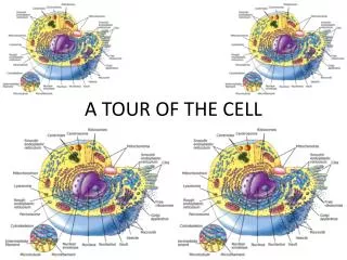
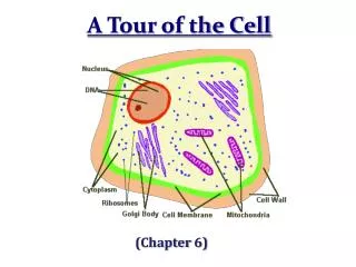
NUCLEUS: An Animal Cell • Nuclear envelope • Chromosomes • Smooth endoplasmic • reticulum • Nucleolus • Rough • endoplasmic • reticulum • Lysosome • Centriole • Ribosomes • Peroxisome • Golgi • apparatus • CYTOSKELETON: • Microtubule • Plasma membrane • Intermediate • filament • Mitochondrion • Microfilament
Cell Membranes • The plasma membrane controls the cell’s contact with the environment • The cytoplasm contains organelles • Many organelles have membranes as boundaries • These compartmentalize the interior of the cell • This allows the cell to carry out a variety of activities simultaneously • Increase total membrane area of the cell
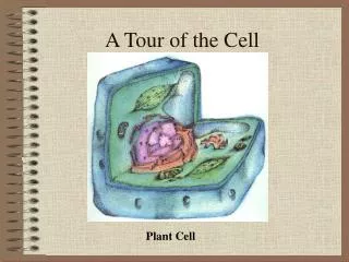
A Plant Cell • All of the membrane-bound organelles present in animal cells are also in plant cells except the lysosome • No centriole or flagellum • Plant cells have structures that animal cells do not have: • Cell wall- protect cells and maintain shape • Chloroplasts – where photosynthesis occurs • Large central vacuole – carry out cellular digestion
Rough endoplasmic • reticulum • NUCLEUS: • Nuclear envelope • Chromosome • Ribosomes • Nucleolus • Smooth • endoplasmic • reticulum • Golgi • apparatus • CYTOSKELETON: • Central vacuole • Microtubule • Chloroplast • Intermediate • filament • Cell wall • Plasmodesmata • Microfilament • Mitochondrion A Plant Cell • Peroxisome • Plasma membrane • Cell wall of • adjacent cell
Hydrophilic head Structure of Membranes • Phosphate • group • Plasma membrane controls movement into and out of the cell • Selective permeability • Structure and component molecules are responsible for this • Made of proteins, some carbohydrates and mainly phospholipids • Symbol • Hydrophobic tails
Structure of Membranes • Phospholipidbilayer • Hydrophilic heads outward, hydophobic tails inward • Impermeable to both hydrophilic and hydrophobic molecules • Proteins attached and embeded in surface layer
Outside cell • Hydrophilic • heads • Hydrophobic • region of • protein • Hydrophobic • tails • Inside cell • Proteins • Hydrophilic • region of • protein
The Nucleus • Genetic control center of a eukaryotic cell • Nuclear DNA is attached to proteins forming very long fibers called chromatin, each fiber constitutes a chromosome • When cell reproduction occurs, the chromosome coils up
The Nucleus • The nucleus is enclosed by a nuclear envelope • Double membrane with pores to control in and outflow of material into the cell • Nucleolus- within the nucleus; mass of fibers and granules; where the components of ribosomes are made
The Nucleus • Two membranes of nuclear envelope • Nucleus • Nucleolus • Chromatin • Pore • Endoplasmic • reticulum • Ribosomes
Ribosomes • Involved in protein synthesis • Synthesized by nucleolus in nucleus • Some ribosomes are free and some are bound • Suspended in cytoplasm • Attached to endoplasmic reticulum
Ribosomes • Ribosomes • Cytoplasm • ER • Endoplasmic reticulum (ER) • Free ribosomes • Bound ribosomes • Large subunit • Small subunit • TEM showing ER and ribosomes • Diagram of a ribosome
Endomembrane System • A biological membrane system in organelles that runs throughout the cell • Some of the membranes are connected, some are not • Many organelles work together in the synthesis, storage, and export of important molecules • Rough and smooth endoplasmic reticulum, the golgi aparatus, lysosomes, and vacuoles
Endoplasmic Reticulum • Two kinds of ER: • Rough ER and smooth ER • These two types of organelles differ in structure and function, but a continuous membrane runs between them • Membranes of rough ER are continuous with the plasma membrane • Space within the ER is separated from the cytoplasm
Transport vesiclebuds off 4 Ribosome Secretory(glyco-) proteininside transportvesicle Sugarchain 3 Glycoprotein 1 2 ROUGH ER Polypeptide Rough ER • Refers to the appearance of this organelle in electron micrographs • Roughness results from ribosomes which stud the membranes of the organelle • Two main functions: • Make more membrane • Make proteins that are secreted by the cell
Smooth ER • Continuous with rough ER • Lacks ribosomes embedded in the membrane • Activity results from enzymes embedded in the membrane • Synthesizes lipids (fatty acids, phospholipids, and steroids) • In some cells (liver), it regulates carbohydrate metabolism and breaks down toxins and drugs
SMOOTH ER ROUGHER Nuclearenvelope Ribosomes SMOOTH ER ROUGH ER Smooth ER • Drug Tolerance • When Liver cells respond to certain drugs over and over they make more ER and become tolerant to the drug • Becomes resistant to the drug and its relatives
Golgi Apparatus • Unconnected, flattened stacks • # of golgi sacks correlates with how active the cell is at secreting proteins • Works with ER by receiving and modifying substances manufactured by the ER • Modifies them and marks them for their destination • The golgi vesicles then bud off and the molecules are shipped to the plasma membrane
Golgi apparatus Golgiapparatus “Receiving” side ofGolgi apparatus Transportvesiclefrom ER Newvesicleforming “Shipping”side of Golgiapparatus Transport vesiclefrom the Golgi Figure 4.10 Golgi Apparatus
Lysosomes • Digestive enzymes enclosed in a membranous sac • Come from the golgi apparatus • Compartmentalize digestive enzymes so they won’t harm the cell • Digest food vacuoles in order to digest them • Destroy harmful bacteria • Recycle damaged organelles • Play important roles in embryonic development
Rough ER Transport vesicle(containing inactivehydrolytic enzymes) Plasmamembrane Golgiapparatus LYSOSOME Engulfmentof particle Lysosomeengulfingdamagedorganelle Nucleus “Food” LYSOSOMES Digestion Foodvacuole Lysozymes
Vacuoles • Membranous sacs that have a variety of functions • Food and chemical storage • Central vacuole of plants aids in plant growth, and chemical and waste storage • Also contain pigments for flower color and poisons that protect plants from predation • In protists they collect water to prevent dilution
Nucleus Centralvacuole Contractilevacuoles Nucleus Vacuoles Protists Plants
Nucleus Nuclear membrane Rough ER Smooth ER Transport vesicle Transport vesicle Lysosome Vacuole Golgi apparatus Plasma membrane
Chloroplasts • The photosynthesizing organelles of plants and protists • Most of the living world runs on the energy provided by photosynthesis • Internal membranes partition the chloroplast into 3 compartments • Intermembrane space • Stroma • Granum
Chloroplast Stroma Inner and outer membranes Granum Intermembranespace Figure 4.15 Chloroplasts • Intermembrane space- between outer and inner membrane of the chloroplast • Stroma- Network of tubules and interconnected hollow disks formed of membranes • Granum- Stacks of hollow disks that are the chloroplasts solarpower packs
Mitochondria • Organelles that convert energy from one chemical form to another • Carry out cellular respiration • Sugars are converted to the chemical energy of a molecule of ATP • Enclosed by two membranes • Composed of two compartments: • Intermembrane space • Mitochondrial matrix
MITOCHONDRION Outermembrane Intermembranespace Innermembrane Cristae Matrix Mitochondria • Intermembrane space • Fluid-filled compartment • Encloses mitochondrial matrix • Mitochondrial matrix- • Many of the rxns of cellular respiration occur here • Inner membrane highly folded into christae that increase the surface area • Embedded in the cristae are the enzymes that make ATP
Cytoskeleton • Cytoskeleton- A supportive meshwork of fine fibers contained in eukaryotic cells • Fibers extend throughout the cell • Also involved in cell movement • May help regulate cellular activity by transmitting signals from the cells exterior to the interior • Three main types of fibers: • Microfiliments, intermediate filiments, microtubules
MICROFILAMENT Actin subunit 7 nm INTERMEDIATE FILAMENT Fibrous subunits 10 nm Cytoskeleton • Microfilaments • Solid helical rods composed mainly of actin • Twisted double chain • Can help cells change shape by assembling and disassembling • Interact with other protein filaments to make cells contract • Intermediate filaments • Fibrous proteins with a rope-like structure • Reinforcing rods and anchor organelles
MICROTUBULE Tubulinsubunit 25 nm Cytoskeleton • Microtubules- straight, hollow tubes composed of globular proteins called tubulin • May disassemble and reassemble to provide rigidity and shape to the cell • Anchorage for organelles and tracks for organelle movement within the cytoplasm
Cilia and Flagella • Cilia- short, numerous appendages • Flagella- longer, less numerous appendages • Core of microtubules wrapped in an extension of the plasma membrane • 9 microtubule doublets surrounds a central pair (9+2) • At the base microtubules form an anchoring structure called the basal body where the central core mt’s disappear • Cilia and flagella provide support and contribute to movement
FLAGELLUM Electron micrograph of sections: Outer microtubule doublet Plasmamembrane Flagellum Centralmicrotubules Outer microtubule doublet Plasmamembrane Basal body Basal body(structurally identical to centriole)
Microtubule doublet Slidingforce Dynein arm Locomotion with Cilia and Flagella • Protein knobs (dynein arms) attached to each microtubule doublet • Bending is done by the dynein arms grabbing onto the adjacent mt and ‘walking’ along it so that the arms slide along each other
Cell surfaces • Most cells have an additional surface coating surrounding the plasma membrane • Prokaryotes-capsules • Interact mainly with non-cellular surroundings • Plants- cell walls • Protect cells and provide structural support • Eukaryotes- composed of many cells organized into a single, functional organism
Plant cell walls • Consist of fibers of the polysaccharide cellulose embedded in a matrix of other polysaccharides and proteins • Cell walls are multi-layered • Between the walls of adjacent cells is a layer of sticky polysaccharides that bonds the cells together