Download
1 / 90
900 likes | 907 Views
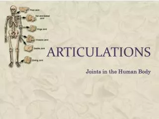
9 - Lecture Articulations. An Introduction to Articulations. Learning Outcomes 9-1 Contrast the major categories of joints, and explain the relationship between structure and function for each category.

E N D
9 - Lecture Articulations
An Introduction to Articulations • Learning Outcomes • 9-1 Contrast the major categories of joints, and explain the relationship between structure and function for each category. • 9-2 Describe the basic structure of a synovial joint, and describe common synovial joint accessory structures and their functions. • 9-3 Describe how the anatomical and functional properties of synovial joints permit movements of the skeleton. • 9-4 Describe the articulations between the vertebrae of the vertebral column.
An Introduction to Articulations • Learning Outcomes • 9-5 Describe the structure and function of the shoulder joint and the elbow joint. • 9-6 Describe the structure and function of the hip joint and the knee joint. • 9-7 Describe the effects of aging on articulations, and discuss the most common age-related clinical problems for articulations. • 9-8 Explain the functional relationships between the skeletal system and other body systems.
An Introduction to Articulations • Articulations • Body movement occurs at joints (articulations) where two bones connect • Joint Structure • Determines direction and distance of movement (range of motion or ROM) • Joint strength decreases as mobility increases
9-1 Classification of Joints • Two Methods of Classification • Functional classification is based on range of motion of the joint • Structural classificationrelies on the anatomical organization of the joint
9-1 Classification of Joints • Functional Classifications • Synarthrosis (immovable joint) • Amphiarthrosis (slightly movable joint) • Diarthrosis (freely movable joint)
9-1 Classification of Joints • Structural Classifications • Bony • Fibrous • Cartilaginous • Synovial
Table 9-1 Functional and Structural Classifications of Articulations
Table 9-1 Functional and Structural Classifications of Articulations
Table 9-1 Functional and Structural Classifications of Articulations
9-1 Classification of Joints • Synarthroses (Immovable Joints) • Are very strong • Edges of bones may touch or interlock • Four types of synarthrotic joints • Suture • Gomphosis • Synchondrosis • Synostosis
9-1 Classification of Joints • Suture • Bones interlocked • Are bound by dense fibrous connective tissue • Are found only in skull • Gomphosis • Fibrous connection (periodontal ligament) • Binds teeth to sockets
9-1 Classification of Joints • Synchondrosis • Is a rigid cartilaginous bridge between two bones • Epiphyseal cartilage of long bones • Between vertebrosternal ribs and sternum • Synostosis • Fused bones, immovable • Metopic suture of skull • Epiphyseal lines of long bones
9-1 Classification of Joints • Amphiarthroses • More movable than synarthrosis • Stronger than freely movable joint • Two types of amphiarthroses • Syndesmosis • Bones connected by ligaments • Symphysis • Bones separated by fibrocartilage
9-1 Classification of Joints • Synovial Joints (Diarthroses) • Also called movable joints • At ends of long bones • Within articular capsules • Lined with synovial membrane
9-2 Synovial Joints • Articular Cartilages • Pad articulating surfaces within articular capsules • Prevent bones from touching • Smooth surfaces lubricated by synovial fluid • Reduce friction
9-2 Synovial Joints • Synovial Fluid • Contains slippery proteoglycans secreted by fibroblasts • Functions of synovial fluid • Lubrication • Nutrient distribution • Shock absorption
9-2 Synovial Joints • Accessory Structures • Cartilages • Fat pads • Ligaments • Tendons • Bursae
9-2 Synovial Joints • Cartilages • Cushion the joint • Fibrocartilage pad called a meniscus (or articular disc; plural, menisci) • Fat Pads • Superficial to the joint capsule • Protect articular cartilages • Ligaments • Support, strengthen joints • Sprain – ligaments with torn collagen fibers
9-2 Synovial Joints • Tendons • Attach to muscles around joint • Help support joint • Bursae • Singular, bursa, a pouch • Pockets of synovial fluid • Cushion areas where tendons or ligaments rub
9-2 Synovial Joints • Factors That Stabilize Synovial Joints • Prevent injury by limiting range of motion • Collagen fibers (joint capsule, ligaments) • Articulating surfaces and menisci • Other bones, muscles, or fat pads • Tendons of articulating bones
Figure 9-1a The Structure of a Synovial Joint Medullary cavity Spongy bone Periosteum Fibrous joint capsule Synovial membrane Articular cartilages Joint cavity(containingsynovial fluid) Compact bone Synovial joint, sagittal section
Figure 9-1b The Structure of a Synovial Joint Quadriceps tendon Bursa Femur Joint capsule Patella Synovialmembrane Articular cartilage Fat pad Meniscus Patellar ligament Intracapsularligament Joint cavity Tibia Meniscus Knee joint, sagittal section
9-2 Synovial Joints • Injuries • Dislocation (luxation) • Articulating surfaces forced out of position • Subluxation • A partial dislocation • Sprain • Tear in a ligament due to joint being carried through ROM greater than normal but without dislocation or fracture • Strain • Tear in a muscle
9-3 Movements • Three Types of Dynamic Motion • Linear movement (gliding) • Angular movement • Rotation • Planes (Axes) of Dynamic Motion • Monaxial (1 axis) • Biaxial (2 axes) • Triaxial (3 axes)
Figure 9-2 A Simple Model of Articular Movement Gliding movement Angular movement Circumduction Rotation Initial position
9-3 Movements • Types of Movement at Synovial Joints • Terms describe: • Plane or direction of motion • Relationship between structures
9-3 Movements • Types of Movement at Synovial Joints • Gliding Movement • Two surfaces slide past each other • Between carpal or tarsal bones
9-3 Movements • Angular Movement • Flexion • Angular motion • Anterior–posterior plane • Reduces angle between elements • Extension • Angular motion • Anterior–posterior plane • Increases angle between elements
9-3 Movements • Angular Movement • Hyperextension • Angular motion • Extension past anatomical position
Figure 9-3a Angular Movements Extension Flexion Hyperextension Flexion Flexion Hyper-extension Extension Extension Flexion Hyperextension Extension Flexion/extension
9-3 Movements • Angular Movement • Abduction • Angular motion • Frontal plane • Moves away from longitudinal axis • Adduction • Angular motion • Frontal plane • Moves toward longitudinal axis
Figure 9-3b Angular Movements Abduction Abduction Adduction Adduction Abduction Adduction Abduction Adduction Abduction/adduction
Figure 9-3c Angular Movements Abduction Adduction Adduction/abduction
9-3 Movements • Angular Movement • Circumduction • Circular motion without rotation • Angular motion
9-3 Movements • Types of Movement at Synovial Joints • Rotation • Direction of rotation from anatomical position • Relative to longitudinal axis of body • Left or right rotation • Medial rotation (inward rotation) • Rotates toward axis • Lateral rotation (outward rotation) • Rotates away from axis
Figure 9-4a Rotational Movements Head rotation Rightrotation Leftrotation Lateral(external)rotation Medial(internal)rotation
9-3 Movements • Rotation • Pronation • Rotates forearm, radius over ulna • Supination • Forearm in anatomical position
9-3 Movements • Special Movements • Inversion • Twists sole of foot medially • Eversion • Twists sole of foot laterally • Dorsiflexion • Flexion at ankle (lifting toes) • Plantar flexion • Extension at ankle (pointing toes) Dorsiflexion(ankle flexion) Plantarflexion(ankle extension)
9-3 Movements Opposition • Opposition • Thumb movement toward fingers or palm (grasping) • Reposition • Opposite of opposition • Protraction • Moves anteriorly • In the horizontal plane (pushing forward) • Retraction • Opposite of protraction • Moving anteriorly (pulling back) Retraction Protraction
9-3 Movements • Special Movements • Elevation • Moves in superior direction (up) • Depression • Moves in inferior direction (down) • Lateral flexion • Bends vertebral column from side to side
Figure 9-5 Synovial Joints Depression Elevation
Figure 9-5 Synovial Joints Lateral flexion
9-3 Movements • Classification of Synovial Joints by Shape • Gliding • Hinge • Pivot • Condylar • Saddle • Ball-and-socket
9-3 Movements • Gliding Joints • Flattened or slightly curved faces • Limited motion (nonaxial) • Hinge Joints • Angular motion in a single plane (monaxial) • Pivot Joints • Rotation only (monaxial)
Figure 9-6 Synovial Joints Gliding joint Movement: slight nonaxial or multiaxial Clavicle Examples: • Acromioclavicular and claviculosternal joints Manubrium • Intercarpal and intertarsal joints • Vertebrocostal joints • Sacro-iliac joints
Figure 9-6 Synovial Joints Hinge joint Movement: monaxial Examples: Humerus • Elbow joint • Knee joint • Ankle joint Ulna • Interphalangeal joint
Figure 9-6 Synovial Joints Pivot joint Movement: monaxial (rotation) Examples: Atlas • Atlanto-axial joint • Proximal radio-ulnar joint Axis
9-3 Movements • Condylar Joints • Oval articular face within a depression • Motion in two planes (biaxial) • Saddle Joints • Two concave, straddled (biaxial) • Ball-and-socket Joints • Round articular face in a depression (triaxial)
Figure 9-6 Synovial Joints Condylar joint Movement: biaxial Examples: • Radiocarpal joint Scaphoidbone • Metacarpophalangeal joints 2–5 • Metatarsophalangeal joints Ulna