Download

1 / 58
990 likes | 4.1k Views
Lehninger Principles of Biochemistry, Fourth Edition, 2005. Chapter 19 OXIDATIVE PHOSPHORYLATION AND PHOTOPHOSPHORYLATION. 中央研究院 生物化學研究所 曾湘文 博士 June 12, 2007 . 19.1 Electron-Transfer Reactions in Mitochondria. Electrons Are Funneled to Universal Electron Acceptors

E N D
Lehninger Principles of Biochemistry, Fourth Edition, 2005 Chapter 19OXIDATIVE PHOSPHORYLATIONAND PHOTOPHOSPHORYLATION 中央研究院 生物化學研究所 曾湘文 博士 June 12, 2007
19.1 Electron-Transfer Reactions in Mitochondria • Electrons Are Funneled to Universal Electron Acceptors • Electrons Pass through a Series of Membrane-Bound Carriers • Electron Carriers Function in Multienzyme Complexes • The Energy of Electron Transfer Is Efficiently Conserved in a Proton Gradient • Plant Mitochondria Have Alternative Mechanisms for Oxidizing NADH
Biochemical anatomy of a mitochondrion • The convolutions (cristae) of the inner membrane provide a very large surface area. • The inner membrane of a single liver mitochondrion may have more than 10,000 sets of electron-transfer systems (respiratory chains) and ATP synthase molecules, distributed over the membrane surface. • Heart mitochondria, which have more profuse cristae and thus a much larger area of inner membrane, contain more than three times as many sets of electron-transfer systems as liver mitochondria. • The mitochondrial pool of coenzymes and intermediates is functionally separate from the cytosolic pool.
Mitochondrion: • like Gram-negative bacteria, have two membranes • The outer mitochondrial membrane is readily permeable to small molecules (Mr 5,000) and ions, which move freely through transmembrane channels formed by a family of integral membrane proteins called porins. • The inner membrane is impermeable to most small molecules and ions, including protons (H); the only species that cross this membrane do so through specific transporters. The inner membrane bears the components of the respiratory chain and the ATP synthase. • Outer membrane contains porins (< 5000). • Matrix pyruvate deHase complex, TCA enzymes, and fatty acid b-oxidation pathway, and amino acids oxidation
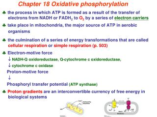
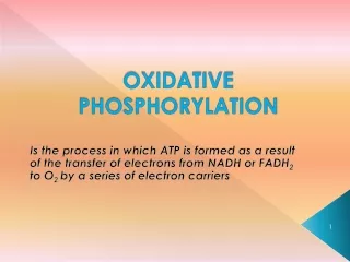
Electrons are funneled to universal electron acceptors • Oxidative phosphorylation begins with the entry of electrons into the respiratory chain. • Most of these electrons arise from the action of dehydrogenases that collect electrons from catabolic pathways and funnel them into universal electron acceptors • nicotinamide nucleotides (NAD or NADP) • flavin nucleotides (FMN or FAD) • Nicotinamide nucleotide–linked dehydrogenases • Flavoproteins
Nicotinamide nucleotide (NAD or NADP)–linked dehydrogenases
Electrons are funneled to universal electron acceptors: NAD and FAD • Most deHase that act in catabolism are specific for NAD+ as electron acceptor • NAD-linked deHase remove two hydrogne atoms from substrates, one of these is transferred as a hydride inon (:H-) to NAD+; the other is releases as H+ in medium. • NADH and NADPH are water-soluble electron carriers that associate reversibly with deHase. NADH carries electrons from catabolic reactions to their point of entry into the ETC, NADPH supplies electrons to anabolic reactions. • Flavoproteins (FMN or FAD) accept either one electron or two.
Flavin nucleotides: FMN or FAD • Flavoproteins (FMN or FAD) accept either one electron (yielding the semiquinone form) or two (yielding FADH2 or FMNH2). • Participate in either one- or two-electron transfers, they can serve as intermediates between reactions in which two electrons are donated (as in dehydrogenations) and those in which only one electron is accepted.
Electrons Pass through a Series of Membrane-Bound Carriers: Uniquinone (Q or coenzyme Q) • Three types of electron transfers occur in oxidative phosphorylation: • direct transfer of electrons, as in the reduction of Fe3+ to Fe2+; • transfer as a hydrogen atom (H+ + e-); and • transfer as a hydride ion (:H-), which bears two electrons. • The term reducing equivalentis used to designate a single electron equivalent transferred in an oxidation-reduction reaction. • Complete reduction of uniquinone requires two electrons and two protons, and occurs in two steps through the semiquinone radical intermediate. • Q is lipid-soluble benzoquinone with a long isoprenoid side chain---freely diffusible within the lipid bilayer of the inner mitochondrial membrane and can shuttle reducing equivalents between other, less mobile electron carriers in the membrane, plays a central role in coupling electron flow to proton movement
Uniquinone (Q or coenzyme Q) • Complete reduction of ubiquinone requires two electrons and two protons, and occurs in two steps through the semiquinone radical intermediate. • a lipid-soluble benzoquinone with a long isoprenoid side chain. • act at the junction between a two-electron donor and a one-electron acceptor. • plastoquinone (in plant chloroplasts) and menaquinone (in bacteria) long isoprenoid side chain
covalently attached through Cys not covalently bound Prosthetic groups of cytochromes • Each group consists of four five-membered, nitrogen-containing rings in a cyclic structure called a porphyrin. • 4 Ns are coordinated with a central Fe ion. • The heme cofactors of aand b cytochromes are tightly, but not covalently; the hemes ofc-type cytochromes are covalently attached through Cys residues. • The cytochromes of type aandband some of type care integral proteins of the inner mitochondrial membrane. • One striking exception is the cytochrome cof mitochondria, a soluble protein that associates through electrostatic interactions with the outer surface of the inner membrane.
Absorption spectra of Cytochrome c • Mitochondria contain three classes of cytochromes, designated a, b, and c, which are distinguished by differences in their light-absorption spectra. Each type of cytochrome in its reduced (Fe2+) state has three absorption bands in the visible range.
Iron-sulfur (Fe-S) proteins/centers • the iron is in association with inorganic sulfur atoms or with the sulfur atoms of Cys residues in the protein, or both. These iron-sulfur (Fe-S) centers range from simple structures with a single Fe atom coordinated to four Cys -SH groups to more complex Fe-S centers with two or four Fe atoms; (a) single Fe, (b) 2Fe-2S, or (c) 4Fe-4S centers. • Rieske ion-sulfur protein (His not Cys) • Participate in one-electron transfers in which one iron atom of the iron-sulfur cluster is oxidized or reduced. • At least 8 Fe-S proteins in ETC
Electrons tend to flow from carriers of lower E’o to higher E’o • Standard reduction potentials: the carriers to function in order of increasing reduction potential, because electrons tend to flow spontaneously from carriers of lower E’o to carriers of higher E’o • however, that the order of standard reduction potentials is not necessarily the same as the order of actual reduction potentials under cellular conditions, which depend on the concentration of reduced and oxidized forms NADH →Q → cytochrome b → cytochrome c1 → cytochrome c → cytochrome a → cytochrome a3 → O2
Method for determining the sequence of electron carriers- inhibitors of electron transfer • A second method for determining the sequence of electron carriers involves reducing the entire chain of carriers experimentally by providing an electron source but no electron acceptor (no O2). • When O2 is suddenly introduced into the system, The carrier nearest O2 (at the end of the chain) gives up its electrons first, the second carrier from the end is oxidized next, and so on. • The actual order depends on concentration of reduced and oxidized forms. • In the presence of O2 and an electron donor, carriers that function before the inhibited step become fully reduced, and those that function after this step are completely oxidized.
Separation of functional complexes of the respiratory chain • The outer mitochondrial membrane is first removed by treatment with the detergent digitonin. • Fragments of inner membrane are then obtained by osmotic rupture of the mitochondria, and the fragments are gently dissolved in a second detergent. • The resulting mixture of inner membrane proteins is resolved by ion-exchange chromatography into different complexes (I through IV) of the respiratory chain, each with its unique protein composition • Complexes I and II catalyze electron transfer to ubiquinone from two different electron donors: NADH (Complex I) and succinate (Complex II). • Complex III carries electrons from reduced ubiquinone to cytochrome c, • Complex IV completes the sequence by transferring electrons from cytochrome c to O2 • Complex V: ATP synthase.
Complex I: NADH to Ubiquinone (NADH: ubiquinone oxidoreductase or NADH dehydrogenase) Complex II: Succinate to Ubiquinone (succinate dehydrogenase) Complex III: Ubiquinone to Cytochrome c (cytochrome bc1 complex or ubiquinone:cytochrome c oxidoreductase,) Complex IV: Cytochrome c to O2 (cytochrome oxidase) Complex V: ATP synthase
Path of electrons from NADH, succinate, fatty acyl-CoA and glycerol 3-phosphate to uniquinone • Electrons from NADH pass through a flavoprotein to a series of iron-sulfur proteins (in Complex I) and then to Q. • Electrons from succinate pass through a flavoprotein and several Fe-S centers (in Complex II) on the way to Q. • Glycerol 3-phosphate donates electrons to a flavoprotein (glycerol 3-phosphate dehydrogenase) on the outer face of the inner mitochondrial membrane, from which they pass to Q. • Acyl-CoA dehydrogenase (the first enzyme of boxidation) transfers electrons from fatty acyl-CoA to electrontransferring flavoprotein (ETF), from which they pass to Q via ETF:ubiquinone oxidoreductase
1.VLCAD (12-18C) 2. MCAD (4-14C) 3. SCAD (4-8C) Trifunctional protein (TFP) inhibit when [NADH]/[NAD]↑ ratio [acetyl-CoA] ↑ The b-Oxidation of Saturated Fatty Acids Has Four Basic Steps • three isozymes of acyl-CoA dehydrogenase-FAD, each specific for a range of fatty-acyl chain lengths: very-long-chain acyl-CoA dehydrogenase: (1).Very-Long-Chain Acyl-CoA Dehydrogenase (VLCAD) - 12 to 18C; (2). MCAD - 4 to 14C; (3). SCAD - 4 to 8C. • enoyl-CoA hydratase: water is added to the double bond of the trans-2-enoyl-CoA to form the L stereoisomer of b-hydroxyacyl-CoA • L-b-hydroxyacyl-CoA is dehydrogenatedto form -ketoacyl-CoA, by the action of – b-hydroxyacyl-CoA dehydrogenase - NAD (inhibit by high [NADH]/[NAD] ratio). • thiolysis:acyl-CoA acetyltransferase (thiolase) promotes reaction of - ketoacyl-CoA with a molecule of free coenzyme A to split off the carboxyl-terminal two-carbon fragment of the original fatty acid as acetyl-CoA. The other product is the coenzyme A thioester of the fatty acid, now shortened by two carbon atoms. acetyl-CoA feedback inhibit thiolase
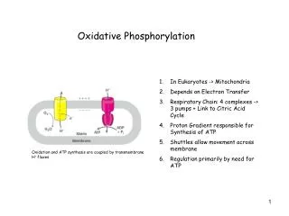
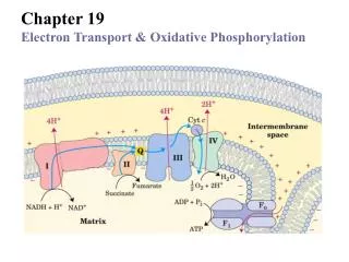
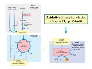
The flow to electrons and protons through the four complexes of the respiratory chain • Electrons reach Q through Complexes I and II. QH2 serves as a mobile carrier of electrons and protons. It passes electrons to Complex III, which passes them to another mobile connecting link, cytochrome c. Complex IV then transfers electrons from reduced cytochrome c to O2. • Electron flow through Complexes I, III, and IV is accompanied by proton flow from the matrix to the intermembrane space. electrons fromoxidation of fatty acids can also enter the respiratory chain through Q. • Much of this energy is used to pump protons out of the matrix. For each pair of electrons transferred to O2, four protons are pumped out by Complex I, four by Complex III, and two by Complex IV.
Complex I - NADH: ubiquinone oxidoreductase vectorial • Complex I catalyzes the transfer of a hydride ion from NADH to FMN, from which two electrons pass through a series of Fe-S centers to the iron sulfur protein N-2 in the matrix arm of the complex. Electron transfer from N-2 to ubiquinone on the membrane arm forms QH2, which diffuses into the lipid bilayer. • This electron transfer also drives the expulsion from the matrix of four protons per pair of electrons. • Blocked by amytal and rotenone.
Complex II: succinate dehydrogenase - Succinate to Ubiquinone • succinate dehydrogenase, the only membrane-bound enzyme in the citric acid cycle. • Electrons move from succinate to FAD, then through the three Fe-S centers to ubiquinone. The heme b is not on the main path of electron transfer but protects against the formation of reactive oxygen species (ROS) hydrogen peroxide (H2O2) and the superoxide radical (O2) that go astray. • Humans with point mutations in Complex II subunits near heme b or the quinone-binding site suffer from hereditary paraganglioma副神經節瘤 . This inherited condition is characterized by benign tumors of the head and neck, commonly in the carotid body頸動脈體 , an organ that senses O2 levels in the blood.
Complex III: cytochromebc1 complex • The complex is a dimer of identical monomers, each with 11 different subunits. • three subunits: cytochrome b (green) with its two hemes (bH and bL, light red); the Rieske iron-sulfur protein (purple) with its 2Fe-2S centers (yellow); and cytochrome c1 (blue) with its heme (red) • The dimeric functional unit. • Blocked by amtimycin.
QP QN The Q cycle • The dimeric functional unit. Cytochrome c1 and the Rieske iron-sulfur protein project from the P surface and can interact with cytochrome c in the intermembrane space. The complex has two distinct binding sites for ubiquinone, QN and QP, • The Q cycle: On the P side of the membrane, two molecules of QH2 are oxidized to Q near the P side, releasing two protons per Q (four protons in all) into the intermembrane space. Each QH2 donates one electron (via the Rieske Fe-S center) to cytochrome c1, and one electron (via cytochrome b) to a molecule of Q near the N side, reducing it in two steps to QH2. This reduction also uses two protons per Q, which are taken up from the matrix.
Complex IV: cytochrome oxidase • the final step of the respiratory chain, carries electrons from cytochrome c to molecular oxygen, reducing it to H2O. • The three proteins critical to electron flow are subunits I, II, and III. The larger green structure includes the other ten proteins in the complex. • Electron transfer through Complex IV begins when two molecules of reduced cytochrome c (top) each donate an electron to the binuclear center CuA. From here electrons pass through heme a to the Fe-Cu center (cytochrome a3 and CuB). • Oxygen now binds to heme a3 and is reduced to its peroxy derivative (O22-) by two electrons from the Fe-Cu center. Delivery of two more electrons from cytochrome c (making four electrons in all) converts the O2 to two molecules of water, with consumption of four “substrate” protons from the matrix. At the same time, four more protons are pumped from the matrix. • Blocked by cyanide anion (CN-), CO, and sodium azide.
The flow to electrons and protons through the four complexes of the respiratory chain • Electrons reach Q through Complexes I and II. QH2 serves as a mobile carrier of electrons and protons. It passes electrons to Complex III, which passes them to another mobile connecting link, cytochrome c. Complex IV then transfers electrons from reduced cytochrome c to O2. • Electron flow through Complexes I, III, and IV is accompanied by proton flow from the matrix to the intermembrane space. electrons fromoxidation of fatty acids can also enter the respiratory chain through Q. • Much of this energy is used to pump protons out of the matrix. For each pair of electrons transferred to O2, four protons are pumped out by Complex I, four by Complex III, and two by Complex IV.
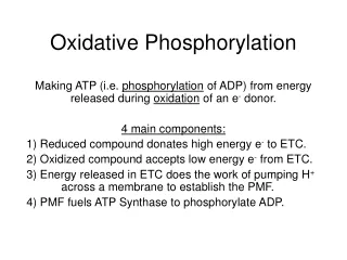
Proton-motive force (pmf) • The energy of electron transfer is efficiently conserved in a proton gradient • The chemical potential energy due to the difference in concentration of H+, and electrical potential energy that results from the separation of charge • The inner mitochondrial membrane separates two compartments of different [H+], resulting in differences in chemical concentration (ΔpH) and charge distribution (Δψ) across the membrane. • The net effect is the proton-motive force (ΔG).
Plant Mitochondria Have Alternative Mechanisms for Oxidizing NADH • Electron carriers of the inner membrane of plant mitochondria. Electrons can flow through Complexes I, III, and IV, as in animal mitochondria, or through plant-specific alternative carriers by the paths shown with blue arrows. • In this process the energy in NADH is dissipated as heat, which can sometimes be of value to the plant. • Cyanide-resistant NADH oxidation is therefore the hallmark of this unique plant electron-transfer pathway.
19.2 ATP Synthesis • ATP Synthase Has Two Functional Domains, Fo and F1 • ATP Is Stabilized Relative to ADP on the Surface of F1 • The Proton Gradient Drives the Release of ATP from the Enzyme Surface • Each b Subunit of ATP Synthase Can Assume Three Different Conformations • Rotational Catalysis Is Key to the Binding-Change Mechanism for ATP Synthesis • Chemiosmotic Coupling Allows Nonintegral Stoichiometries of O2 Consumption and ATP Synthesis • The Proton-Motive Force Energizes Active Transport • Shuttle Systems Indirectly Convey Cytosolic NADH into Mitochondria for Oxidation
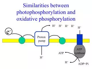
Chemiosmotic model • electrons from NADH and other oxidizable substrates pass through a chain of carriers arranged asymmetrically in the inner membrane. • Electron flow is accompanied by proton transfer across the membrane, producing both a chemical gradient (ΔpH) and an electrical gradient (Δψ). • The inner mitochondrial membrane is impermeable to protons; protons can reenter the matrix only through proton-specific channels (Fo). • The proton-motive force that drives protons back into the matrix provides the energy for ATP synthesis, catalyzed by the F1 complex associated with Fo.
Coupling of electron transfer and ATP synthesis • Chemiosmotic theory readily explains the dependence of electron transfer on ATP synthesis in mitochondria. • Addition of ADP and Pi alone results in little or no increase in either respiration (O2 consumption; black) or ATP synthesis (red). Whensuccinate is added, respiration begins immediately and ATP is synthesized. Addition of cyanide (CN),which blocks electron transfer between cytochrome oxidase and O2, inhibits both respiration and ATP synthesis. • Mitochondria provided with succinate respire and synthesize ATP only when ADP and Pi are added. Subsequent addition of venturicidinoroligomycin, inhibitors of ATP synthase, blocks both ATP synthesis and respiration. • Dinitrophenol (DNP) is an uncoupler, allowing respiration to continue without ATP synthesis.
Chemical uncouplers • Both DNP and FCCP have a dissociable proton and are very hydrophobic. • They carry protons across the inner mitochondrial membrane, dissipating the proton gradient.
Evidence for the role of a proton gradient in ATP synthesis • An artificially imposed electrochemical gradient can drive ATP synthesis in the absence of an oxidizable substrate as electron donor. • (a) isolated mitochondria are first incubated in a pH 9 buffer containing 0.1 M KCl. Slow leakage of buffer and KCl into the mitochondria eventually brings the matrix into equilibrium with the surrounding medium. No oxidizable substrates are present. • (b) Mitochondria are now separated from the pH 9 buffer and resuspended in pH 7 buffer containing valinomycin (K+ ionophore) but no KCl. The change in buffer creates a difference of two pH units across the inner mitochondrial membrane. The outward flow of K, carried (by valinomycin) down its concentration gradient without a counterion, creates a charge imbalance across the membrane (matrix negative). • The sum of the chemical potential provided by the pH difference and the electrical potential provided by the separation of charges is a proton motive force large enough to support ATP synthesis in the absence of an oxidizable substrate.
Mitochondrial ATP synthase complex (FoF1 complex) • The two b subunits of Fo associate firmly with the aand bsubunits of F1, holding them fixed relative to the membrane. • In Fo, the membrane-embedded cylinder of c subunits is attached to the shaft made up of F1 subunits and . • As protons flow through the membrane from the P side to the N side through Fo, the cylinder and shaft rotate, and the subunits of F1 change conformation as the subunit associates with each in turn.
Rotational Catalysis Is Key to the Binding-Change Mechanism for ATP Synthesis • The F1 complex has three nonequivalent adenine nucleotide–binding sites, one for each pair of aand bsubunits. • At any given moment, one of these sites is in theb-ATPconformation (which binds ATP tightly), a second is in theb-ADP (loose-binding) conformation, and a third is in the b-empty (very-loose-binding) conformation. • The proton-motive force causes rotation of the central shaft—theg subunit, which comes into contact with each subunit pair in succession. This produces a cooperative conformational change in which the b-ATP site is converted to the b-empty conformation, and ATP dissociates; the b-ADP site is converted to the b-ATP conformation, which promotes condensation of bound ADP Pi to form ATP; and the -empty site becomes a –b-ADP site, which loosely binds ADP Pi entering from the solvent. • This model, based on experimental findings, requires that at least two of the three catalytic sites alternate in activity; ATP cannot be released from one site unless and until ADP and Pi are bound at the other.
Rotation of Fo and g-subunit • F1 genetically engineered to contain a run of His residues adheres tightly to a microscope slide coated with a Ni complex; biotin is covalently attached to a c subunit of Fo. The protein avidin, which binds biotin very tightly, is covalently attached to long filaments of actin labeled with a fluorescent probe. Biotin-avidin binding now attaches the actin filaments to the c subunit. • When ATP is provided as substrate for the ATPase activity of F1, the labeled filament is seen to rotate continuously in one direction, proving that the Fo cylinder of c subunits rotates. • In another experiment, a fluorescent actin filament was attached directly to the subunit. The series of fluorescence micrographs shows the position of the actin filament at intervals of 133 ms. • Note that as the filament rotates, it makes a discrete jump about every eleventh frame. Presumably the cylinder and shaft move as one unit.
Adenine nucleotide and phosphate translocases • ATP synthasome • Transport systems of the inner mitochondrial membrane carry ADP and Pi into the matrix and newly synthesized ATP into the cytosol. • Adenine nucleotide translocase is an antiporter; the same protein moves ADP into the matrix and ATP out. The effect of replacing ATP4- with ADP3- is the net efflux of one negative charge, which is favored by the charge difference across the inner membrane (outside positive). At pH 7, Pi is present as both HPO42- and H2PO4-; • Phosphate translocase is specific for H2PO4-. There is no net flow of charge during symport of H2PO4- and H+, but the relatively low proton concentration in the matrix favors the inward movement of H+. • Proton-motive force (pmf) is responsible both for providing the energy for ATP synthesis and for transporting substrates (ADP and Pi) in and product (ATP) out of the mitochondrial matrix.
a-ketoglutarate Glutamate Malate-Aspartate Shuttle (liver, kidney, heart)
Malate-Aspartate Shuttle (liver, kidney, heart) • NADH in the cytosol (intermembrane space) passes two reducing equivalents to oxaloacetate, producing malate. • Malate crosses the inner membrane via the malate-a-ketoglutarate transporter. • In the matrix, malate passes two reducing equivalents to NAD+, and the resulting NADH is oxidized by the respiratory chain. • The oxaloacetate formed from malate cannot pass directly into the cytosol. It is first transaminated to aspartate, • Asparate can leave via the glutamate-aspartate transporter. • Oxaloacetate is regenerated in the cytosol, completing the cycle. • NADH can pass electrons directly to the respiratory chain of complex I. About 2.5 molecules of ATP are generated as this pair of electrons passes to O2.
Glycerol 3-phosphate shuttle (Skeletal muscle and brain) • Skeletal muscle and brain use a different NADH shuttle, the glycerol 3-phosphate shuttle. • It differs from the malate-aspartate shuttle in that it delivers the reducing equivalents from NADH to ubiquinone and thus into Complex III, not Complex I providing only enough energy to synthesize 1.5 ATP molecules per pair of electrons. • not involve membrane transport systems.
19.3 Regulation of Oxidative Phosphorylation • Oxidative Phosphorylation Is Regulated by Cellular Energy Needs • An Inhibitory Protein Prevents ATP Hydrolysis during Ischemia • Uncoupled Mitochondria in Brown Fat Produce Heat • ATP-Producing Pathways Are Coordinately Regulated
Oxidative Phosphorylation Is Regulated by Cellular Energy Needs • The rate of respiration (O2 consumption) is generally limited by the availability of ADP as a substrate for phosphorylation. • In some animal tissues, the acceptor control ratio, the ratio of the maximal rate of ADP-induced O2 consumption to the basal rate in the absence of ADP, is at least ten. • the mass-action ratioof the ATP-ADP system ([ATP]/([ADP][Pi]). • some energy-requiring process (eg. Protein synthesis) increases, the rate of breakdown of ATP to ADP and Pi increases, • lowering the mass-action ratio. • With more ADP available for oxidative phosphorylation, • the rate of respiration increases, causing regeneration of ATP.
An Inhibitory Protein (IF1) Prevents ATP Hydrolysis during Ischemia • When a cell is ischemic (deprived of oxygen), as in a heart attack or stroke, electron transfer to oxygen ceases, and so does the pumping of protons. The proton-motive force soon collapses. • the ATP synthase operate in reverse, hydrolyzing ATP to pump protons outward [ATP]↓ • protein inhibitor, IF1, a small (84 amino acids), binds to two ATP synthase and inhibit their ATPase activity • IF1 is inhibitory only in its dimeric form, which is favored at pH lower than 6.5. In a cell starved for oxygen, the main source of ATP becomes glycolysis, and the pyruvic or lactic acid thus formed lowers the pH in the cytosol and the mitochondrial matrix. This favors IF1 dimerization, leading to inhibition of the ATPase • In aerobic metabolism resumes, production of pyruvic acid slows, the pH of the cytosol rises, the IF1 dimer is destabilized, and the inhibition of ATP synthase is lifted.
Heat generation by uncoupled mitochondria • Most newborn mammals, brown fatfuels oxidation serves not to produce ATP but to generate heat to keep the newborn warm. • Brown fat with large numbers of mitochondria and thus large amounts of cytochromes, whose heme groups are strong absorbers of visible light. • Thermogenin(uncoupling protein) short-circuiting of protons, the energy of oxidation is not conserved by ATP formation but is dissipated as heat – maintaining the body temperature of the newborn. • Hibernating animals also depend on uncoupled mitochondria of brown fat to generate heat during their long dormancy
Regulation of The ATP-production pathways • acceptor control ratio: the ratio of the maximal rate of ADP-induced O2 consumption to the basal rate in the absence of ADP. • ATP-producing pathways are coordinately regulated. • Interlocking regulation of glycolysis, pyruvated oxidation, the citric acid cycle ,and oxidation phosphorylation by the relative conc. of ATP, ADP, and AMP, and by NADH. • All four pathway are accelerate when the use of ATP and the formation of ADP, AMP, and Pi increase. • Interlocking of glycolysis and the citric acid cycle by citrate, which inhibits glycolysis, supplements the action of the adenine nucleotide system • Increase NADH and acetyl-CoA inhibit the oxidation of pyruvate to acetyl-CoA, • High NADH/NAD+ ratios inhibit the dehydrogenase reaction of TCA cycle.
Mitochondrial genes and mutations • Mitochondria disease--maternal transmitted —LHON (leber’s hereditary optic遺傳性視神經萎縮症, ND4 gene of complex I Arg to His) —NADH to uniquinone is defected not enough ATP for neurons. • Single base change in cytochrome b of complex III---LHON . • Myoclonic epilepsy and ragged red fiber disease (MERRF, 肌攣性癲癇和殘破紅肌纖維病變)—tRNA (leucyl-tRNA)---protein defective-prarcrystalline structure. • The accumulation of mutations in mitochondrial DNA during a lifetime of exposure to DNA-damaging ---not enough ATP. • human mitochondrial genome contains 37 genes (16,569 bp): 13 subunits of ETS proteins and 24 rRNA and tRNA gene 900 mitochondrial proteins are encoded by nuclear - synthesized in cytosol then imported and assembled within the mitochondria