Download
1 / 29
550 likes | 3.07k Views
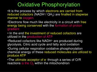
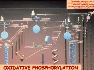
Chapter 18 Oxidative phosphorylation. the process in which ATP is formed as a result of the transfer of electrons from NADH or FADH 2 to O 2 by a series of electron carriers take place in mitochondria, the major source of ATP in aerobic organisms

E N D
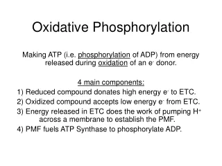
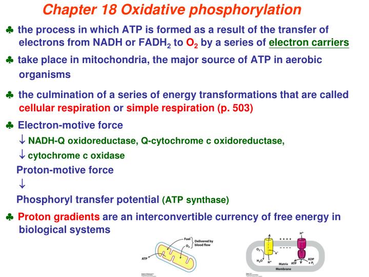
Chapter 18 Oxidative phosphorylation the process in which ATP is formed as a result of the transfer of electrons from NADH or FADH2 to O2 by a series of electron carriers take place in mitochondria, the major source of ATP in aerobic organisms the culmination of a series of energy transformations that are called cellular respiration or simple respiration (p. 503) Electron-motive force NADH-Q oxidoreductase, Q-cytochrome c oxidoreductase, cytochrome c oxidase Proton-motive force Phosphoryl transfer potential (ATP synthase) Proton gradients are an interconvertible currency of free energy in biological systems
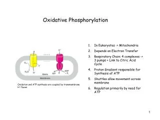
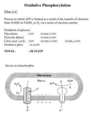
§18.1Oxidative phosphorylation in eukaryotes takes place in mitochondria:2 m in length and 0.5 m in diameter Kennedy and Lehninger (TCA cycle, fatty acid oxidation) quite permeable voltage-dependent anion channel (mitochondrial porin) (oxidative phosphorylation) Impermeable a large family of transporters shuttles metabolites matrix side (N side) cytosolic side (P side)
§18.2Oxidative phosphorylationdepends on electron transfer Measurement of redox potential (E0’) to evaluate electron-transfer potential (G°’) 1M reduction potential of H+:H2 couple = 0
G°= -nF E0 faraday (23.05 kcal mol-1V-1) ½ O2 + NADH + H+ H2O + NAD+ G0' = - 52.6 kcal mole-1p. 508 Release energy is used 1. proton gradient formation ATP synthesis ATP hydrolysis G0' = -7.3 kcal mole-1 2. transport metabolites across the Mito. membrane H+matrixcyto: 5.2 kcal mole-1 △ G = RT ln(C2/C1) + ZF △V pH lower
§ 18.3 Four complexes in respiratory chain Respirasome 1,2,3 1,2,4 ? Electron affinity high
Nelson does not pump protons
P N
Respiratory chain complexes separation Nelson ATP synthase (complex V) In vitro, hydrolytic activity
Universal electron acceptors: NADH and NADPH: are water soluble, can’t cross inner Mito. membrane carry e- from catabolic rxs. vs. supply e- to anabolic rxs. [reduced form]/[oxidized form] Nelson hydride UV p. 499
Universal electron acceptors: Flavin nucleotides (FMN or FAD): are bound to flavoproteins which determine the reduction potential of a flavin nucleotide a part of the flavoprotein’s active site flavoproteins can participate in either one- or two- electron transfer Nelson
Nelson Universal electron acceptors: Ubiquinone (coenzyme Q, Q): a lipid-soluble molecule can accept one or two e- carry both e- and proton Q pool: a pool of Q and QH2 exist in the inner Mito. membrane
Nelson Universal electron acceptors: iron-sulfur proteins:one-electron transfer non-heme iron proteins without releasing or binding protons p. 511 1 Fe — 4 Cys 2 Fe — 2 S — 4 Cys 4 Fe — 4 S — 4 Cys Rieske iron-sulfur proteins: 2 His residues replace 2 cys residues Phosphorylation at His
Universal electron acceptors: cytochromes: a, b, c three classes in Mito. one-electron transfer Nelson Covalently associated to proteins Vinyl group 560 nm 550 nm (C17) The standard reduction potential (p. 507) The longest-wavelength 600 nm
Reduced state (Fe2+) Nelson Color?
1. NADH-Q oxidoreductase(NADH dehydrogenase, complex Ⅰ) NADH + Q + 5H+matrix NAD+ + QH2 + 4H+cytosol
2. Succinate-Q reductase (complex Ⅱ) Nelson p. 528
Q cycle: semiquinone radical anion
3. Q-cytochrome c oxidoreductase(cytochrome bc1 complex; cytochrome reductase; complex Ⅲ) His replace cys 1e- 1e- Q 3(hemes)cytochrome c 1(2Fe-2S) during Q cycle
4. Complex Ⅳ: Cytochrome c oxidase e- from cytosol to O2 2 heme a, 3 copper ions 3 subunits CuA/CuA heme a heme a3 CuB O2 ferric/ferrous cupric/cuprous 4 cyt cred +8 H+N + O2 4 cyt cox + 2 H2O + 4 H+P Nelson ?
1st e- Cupric (Cu2+) Cuprous (Cu+) 2nd e- Ferric (Fe3+) Ferrous (Fe2+) 3th and 4th e-
Proton transport by complex Ⅳ4 cyt cred + 8 H+N + O2 4 cyt cox + 2 H2O + 4 H+P G0’ 4 H+ 5.2 kcal/mole(p. 509) 2 23.06 0.82 (Tab. 18.1) Charge neutrality and Conformational changes (p. 517)
only electrons transfer, no protons transport NADH + 11 H+N + ½ O2 → NAD+ + 10 H+p + H2O FADH2 6
Reactive (active) oxygen species (R[A]OSs) Danger lurks in the reduction of O2 superoxide radical (·O2-), peroxide (O22-), hydrogen peroxide (H2O2),hydroxyl radical (OH·),singlet oxygen (O21) superoxide dismutase (SOD): Cu/Zn-; Mn-; Fe- catalase (CAT): 2 H2O2 O2 + 2H2O a heme protein peroxidase: H2O2 + RH2 2 H2O + R [ascorbate or glutathione peroxidase] SOD: 2 ·O2- + 2H+ O2 + H2O2 Dismutation: a reaction in which a single reactant is converted into two different products Antioxidant vitamins: Vit C: Vit E: lipophilic, avoid lipid peroxidation
Radical ·Q– from complex Ⅰto QH2 QH2 to bL of complex III Nelson p. 722 Also from pentose phosphate pathway
TypeⅠ: insulin dep. a paucity of pancreatic cells TypeⅡ: non-insulin dep. slow to develop, in older, obese individuals insulin is produced, but some feature of the insulin-response system is defective The characteristic symptoms of both types: polydipsia, polyuria, glucosuria Aerobic metabolism More ROS More protective enzymes were induced