Download
1 / 99
1.16k likes | 1.67k Views
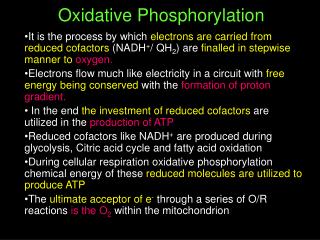
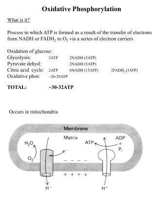
OXIDATIVE PHOSPHORYLATION. Is the process in which ATP is formed as a result of the transfer of electrons from NADH or FADH 2 to O 2 by a series of electron carriers. Cellular respiration. Carbon fuels are oxidized in the citric acid cycle to yield electrons with high transfer potential.

E N D
OXIDATIVE PHOSPHORYLATION Is the process in which ATP is formed as a result of the transfer of electrons from NADH or FADH2 to O2 by a series of electron carriers
Cellular respiration • Carbon fuels are oxidized in the citric acid cycle to yield electrons with high transfer potential. • This electron-motive force is converted into a proton-motive force. • The proton-motive force is converted into phosphoryl transfer potential.
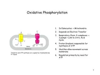
Essence of Oxidative Phosphorylation • Oxidation and ATP synthesis are coupled by transmembrane proton fluxes.
Mitochondria: TWO COMPARTMENTS • The intermembrane space between the outer and the inner membranes • Oxidative phosphorylation • The matrix which is bounded by the inner membrane: • most of the reactions of the citric acid cycle and fatty acid oxidation
Components of the mitochondrial electron-transport chain • Large multi subunit integral membrane protein complexes or coupling sites: couple electron transfer with H+ gradient generation • Complex I:NADH-Q oxidoreductase(MW =880 kd) Coupling Site 1 • Complex II:succinate-Q reductase complex (MW =140 kd) • Complex III:Q-cytochrome c oxido-reductase(MW = 250 kd). Coupling Site 2 • Complex IV:cytochrome c oxidase(MW = 160 kd) Coupling Site 3.
Small Mobile Electron Carriers: • Ubiquinone/Ubiquinol (Q/QH2): small hydrophobic electron carriers which shuttle electrons between the large complexes and back and forth across the lipid bilayer. • Cytochrome c: is a small water soluble protein which is a mobile electron carrier and carries electrons between cytochrome bc1 complex and cytochrome c oxidase.
Electron flow within these transmembrane complexes leads to the transport of protons across the inner mitochondrial membrane. • Electrons are carried from NADH-Q oxidoreductase to Q-cytochrome c oxidoreductase by the reduced form of coenzyme Q (Q). • Q also carries electrons from FADH2, generated in succinate dehydrogenase or (succinate-Q reductase) in the citric acid cycle, to Q-cytochrome c oxidoreductase • Cytochrome c, a small, soluble protein, shuttles electrons from Q-cytochrome c oxidoreductase to cytochrome c oxidase (complex IV), which catalyzes the reduction of O2. • Succinate-Q reductase (Complex II), in contrast with the other complexes, does not pump protons.
Coenzyme Q (Q) • Q is a hydrophobic quinone that diffuses rapidly within the inner mitochondrial membrane. • Q is a quinone derivative with a long isoprenoid tail. • The number of five-carbon isoprene units in coenzyme Q depends on the species. • The most common form in mammals contains 10 isoprene units (coenzyme Q10).
Oxidation States of Quinones The reduction of ubiquinone (Q) to ubiquinol (QH2) proceeds through a semiquinone anion intermediate (Q.-). In the fully oxidized state (Q), coenzyme Q has two keto groups. The addition of one electron and one proton results in the semiquinone form (QH·). The semiquinone form is relatively easily deprotonated to form a semiquinone radical anion (Q·-). The addition of a second electron and proton generates ubiquinol (QH2),
Thus, electron-transfer reactions of quinones are coupled to proton binding and release, a property that is key to transmembrane proton transport.
Flavins • Oxidation States of Flavins: • The reduction of flavin mononucleotide (FMN) to FMNH2 proceeds through a semiquinone intermediate.
Fe-S clusters • Fe-Sclusters in iron-sulfur proteins (nonheme iron proteins) play a critical role in a wide range of reduction reactions in biological systems. • Iron ions in these Fe-S complexes cycle between: • Fe2+ (reduced state) • Fe3+ (oxidized state) • Unlike quinones and flavins, iron-sulfur clusters generally undergo oxidation-reduction reactions without releasing or binding protons.
Several types of Fe-S clusters are known: • A single iron ion bound by four cysteine residues. • 2Fe-2S cluster with iron ions bridged by sulfide ions. • 4Fe-4S cluster. • Each of these clusters can undergo oxidation-reduction reactions.
COMPLEX INADH-Q oxidoreductase • NADH-Q oxidoreductase (also called NADH dehydrogenase): • an enormous enzyme (880 kd) • consists of at least 34 polypeptide chains. • consists of a membrane-spanning part and a long arm that extends into the matrix. • Contains FMN and Fe-S prosthetic groups.
NADH is oxidized in the arm, and the electrons are transferred to reduce Q in the membrane. • The reaction catalyzed by this enzyme appears to be:
FMNH2 FMN NAD+ NADH + H+ Cytosol • The initial step is the binding of NADH and the transfer of its two high-potential electrons to the flavin mononucleotide (FMN) prosthetic group of this complex to give the reduced form, FMNH2. Matrix
Fe2+ Fe3+ FMNH2 FMN NAD+ NADH + H+ 2H+ • Electrons are then transferred from FMNH2 to a series of three 4Fe4S, the second type of prosthetic group in NADH-Q oxidoreductase. • NADH-Q oxidoreductase contains: • 2Fe-2S cluster • 4Fe-4S cluster • Two protons move back to the matrix Matrix
UQH2 UQ Fe2+ Fe3+ FMNH2 FMN NAD+ NADH + H+ 2H+ 2H+ Cytosol • Electrons in the 4Fe4S of NADH-Q oxidoreductase are shuttled to coenzyme Q. • Two hydrogen ions are pumped out of the matrix of the mitochondrion. Matrix
2H+ UQH2 UQ Fe2+ Fe3+ UQH2 UQ Fe2+ Fe3+ FMNH2 FMN NAD+ NADH + H+ 2H+ 2H+ 2H+ Cytosol • The pair of electrons on bound QH2 are transferred to a 4Fe-4S center and the protons are released on the cytosolic side. • These electrons are transferred to a mobile Q in the hydrophobic core of the membrane, resulting in the uptake of two additional protons from the matrix. Matrix
In summary: • NADH binds to a site on the vertical arm and transfers its electrons to FMN. • These electrons flow within the vertical unit to three 4Fe-4S centers. • Then they flow to a bound Q. The reduction of Q to QH2 results in the uptake of two protons from the matrix. • 4 Protons are pumped this way which is still under investigation
COMPLEX IIsuccinate-Q reductase complex • It is an integral membrane protein of the inner mitochondrial membrane. • Contains three different kinds of Fe-S clusters: • 2Fe-2S • 3Fe-3S • 4Fe-4S.
FADH2 is generated in the citric acid cycle by the enzyme succinate dehydrogenase, with the oxidation of succinate to fumarate.
Two electrons are transferred from FADH2 directly to Fe-S clusters of succinate dehydrogenase. • The electrons are then passed to (Q) for entry into the electron-transport chain. • FADH2 is generated by other reactions (such as: Glycerol phosphate dehydrogenase and fatty acyl CoA dehydrogenase). • This FADH2 also transfers its electrons to (Q), to form (QH2).
The succinate-Q reductase complex and other enzymes that transfer electrons from FADH2 to Q, in contrast with NADH-Q oxidoreductase, do not transport protons. • Consequently, less ATP is formed from the oxidation of FADH2 than from NADH.
COMPLEX IIIQ-Cytochrome c Oxidoreductase • The second of the three proton pumps in the respiratory chain (also known as cytochrome reductase). • The function of Q-cytochrome c oxidoreductase is to catalyze the transfer of electrons from QH2to oxidized cytochrome c (cyt c), a water-soluble protein, and concomitantly pump protons out of the mitochondrial matrix.
Structure of Q-CytochromeCOxidoreductase (CytochromeBC1). This enzyme is a homodimer with 11 distinct polypeptide chains. The major prosthetic groups, three hemes and a 2Fe-2S cluster, mediate the electron-transfer reactions between quinones in the membrane and cytochrome c in the intermembrane space.
Structure of Q-CytochromeCOxidoreductase • Q-cytochromecoxidoreductase contains: • Cytochrome b562 • Cytochrome b566 • Cytochrome c1 • An iron sulfur protein • At least six other subunits. • A cytochrome is an electron-transferring protein that contains a heme prosthetic group. • The iron ion of a cytochrome alternates between a reducedferrous (+2) state and an oxidizedferric (+3) state during electron transport.
Structure of Q-CytochromeCOxidoreductase • The enzyme contains three heme prosthetic groups contained within two cytochrome subunits: • Two b-type hemes within cytochrome b: • Heme bL (L for low affinity) • Heme bH (H for high affinity) • One c-type heme within cytochrome c1 (Heme c1). • Hemes in the 3 classes of cytochrome (a, b, c) differ slightly in substituents on the porphyrin ring system. • Only heme c is covalently linked to the protein via thioether bonds to cysteine residues
Structure of Q-CytochromeCOxidoreductase • The enzyme also contains an iron-sulfur protein with an 2Fe-2Scenter (Rieske center). • This center is unusual in that one of the iron ions is coordinated by two his residues rather than two cysteine residues. • This coordination stabilizes the center in its reduced form, raising its reduction potential. • Finally, Q-cytochrome c oxidoreductase contains two distinct binding sites for ubiquinone termed (Qo) and (Qi), with the Qi site lying closer to the inside of the matrix.
The Q Cycle • The mechanism for the coupling of electron transfer from Q to cytochrome c to transmembrane proton transport. • Facilitates the switch from the two-electron carrier ubiquinol to the one-electron carrier cytochrome c.
The first electron: • flows first to the Rieske 2Fe-2S cluster • then to cytochrome c1 • finally to a molecule of oxidized cytochrome c, converting it into its reduced form. • The second electron: • transferred first to cytochrome bL • then to cytochrome bH • finally to an oxidized uniquinone bound in the Qi site. • This quinone (Q) molecule is reduced to a semiquinone anion (Q·-). Cyt c Cyt c1 Fe-S QH2 QH Q Qo site H+ H+ Cyt bL Cyt bH Q Qi site Q.-
Cyt c Cyt c1 Fe-S QH2 Cyt bL Cyt bH Q.- Q Qo site Qi site
QH2 Q.- H+ H+ H+ H+ Cyt c Cyt c1 Fe-S Q QH2 Qo site Cyt bL Cyt bH Qi site
(QH2) binds in the Qo site, and transfers its electrons, one at a time:
COMPLEX IV Cytochrome c Oxidase • It catalyzes the coupled oxidation of the reduced cyt c generated by Complex III, and reduction of O2to two molecules of H2O. • The four-electron reduction of oxygen directly to water without the release of intermediates is quite thermodynamically favorable. DG°´ = -231.8 kJ mol-1 • As much of this free energy as possible must be captured in the form of a proton gradient for subsequent use in ATP synthesis.
It consists of 13 subunits, 3 of which (subunits I, II, and III) are encoded by the mitochondrial genome. • Cytochrome c oxidase contains: • Three copper ions, arranged as two copper centers, designated A and B: • CuA/CuA: contains two copper ions linked by two bridging cysteine residues. This center initially accepts electrons from reduced cytochrome c. • CuB: is coordinated by three histidine residues, one of which is modified by covalent linkage to a tyrosine residue. • Two heme A groups: • Heme a: functions to carry electrons from CuA/CuA • heme a3: passes electrons to CuB, to which it is directly adjacent. • Together, heme a3 and CuB form the active center at which O2 is reduced to H2O.
Cytochrome C Oxidase. • The major prosthetic groups include: • CuA/CuA • heme a • heme a3-CuB. • Heme a3-CuB is the site of the reduction of oxygen to water. .
Cyt c Cyt c FIRST ELECTRONE CuA/CuA Electrone transfer to CuB Heme A Heme a3 CuB
Cyt c Cyt c SECOND ELECTRONE CuA/CuA Electrone transfer to Fe in Heme a3 Heme A Both CuBand Fe in Heme a3are in reduced form Heme a3 O O Binding of O2 CuB Formation of peroxide bridge O O
Cyt c Cyt c THIRD ELECTRONE CuA/CuA Heme A Heme a3 O O CuB Cleavage of O-O bond H+
Cyt c Cyt c FOURTH ELECTRONE CuA/CuA Heme A H+ Heme a3 O O CuB Reduction of the Ferryl group H+
Cyt c CuA/CuA Heme A H+ H+ Heme a3 O O CuB H+ H+ Release of water H+ H+
Cyt c Cyt c H+ H+ O O H+ H+ CuA/CuA Heme A Heme a3 CuB
This reaction can be summarized as • The four protons in this reaction come exclusively from the matrix. • Thus, the consumption of these four protons contributes directly to the proton gradient. • Each proton contributes (21.8 kJ mol-1) to the free energy associated with the proton gradient.
Proton Transport by CytochromeC Oxidase. • Four "chemical" protons are taken up from the matrix side to reduce one molecule of O2 to two molecules of H2O. • Four additional "pumped" protons are transported out of the matrix and released on the Cytosolic side in the course of the reaction. • The pumped protons double the efficiency of free-energy storage in the form of a proton gradient for this final step in the electron-transport chain.
How these protons are transported through the protein? • Still under study. • However, two effects contribute to the mechanism: • Charge neutrality tends to be maintained in the interior of proteins. • Thus, the addition of an electron to a site inside a protein tends to favor the binding of a proton to a nearby site. • Conformational changes take place, particularly around the hemea3-CuB center, in the course of the reaction cycle. • These changes must be used to allow protons to enter the protein exclusively from the matrix side and to exit exclusively to the cytosolic side.
Thus, the overall process catalyzed by cytochrome c oxidase is. • molecular oxygen is an ideal terminal electron acceptor, because its high affinity for electrons provides a large thermodynamic driving force.
The reduction of O2 is safe because: Cytochrome c oxidase does not release partly reduced intermediates by holding O2 tightly between Fe and Cu ions. However, partial reduction generates small amounts of hazardous compounds (ROS). Superoxide anion peroxide. Reactive oxygen species or ROS.