Download

1 / 56
560 likes | 614 Views
CHARACTERISTICS OF PROKARYOTIC AND EUKARYOTIC CELLS. CHAPTER 4.

E N D
CHARACTERISTICS OF PROKARYOTIC AND EUKARYOTIC CELLS CHAPTER 4
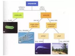
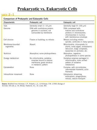
Detailed studies of cells have revealed that prokaryotes differ enough to be split into two large groups called domains. A relatively new concept in classification, domain is the highest taxonomic category, higher even than kingdom. Three domains exist: • Archaea (archaeobacteria) • Bacteria (eubacteria) • Eukarya Archaea and Bacteria are both prokaryotes. The comparison in the next few slides is between bacteria and Eukaryotic cells. We will deal with archaea in a following slide.
Basic Cell Types: Prokaryotic are cells that lack a nucleus (nuclear membrane). Prokarotic cells are single cells but are subdivided into Bacteria and Arachaea as mention in the previous slide. Eukaryotic cells contain a nucleus (nuclear membrane). Eukaryotic cells include: plants, animals, fungi and protists ( a very heterogeneous group that are neither animals, plants or fungi and are often single cell and small e.g., protozoa). Prokaryotes (Bacteria) and Eukaryotes have many similarities and many differences:
Size: Most prokaryotes range from 0.5 to 2.0 µm in diameter. A human red blood cell is about 7.5 µm.
Shape Bacteria which show a wide variety of shapes within a single species are said to be Pleomorphic. Those of you taking the laboratory will see the various shapes of bacteria first-hand. Fig. 4.1 The most common bacterial shapes
Fig. 4.2 Arrangements of some of The more common cocci forms of bacteria
1. Cell membrane-typically surrounded by a cell wall 2. Internal cytoplasm with ribosomes, a nuclear region and granules 3. Lots of external structure, e.g., capsules, flagella and pili Fig. 4.3 A typical prokaryotic cell
1. Cell membrane-typically surrounded by a cell wall 2. Internal cytoplasm with ribosomes, a nuclear region and granules 3. Lots of external structure, e.g., capsules, flagella and pili Fig. 4.3 A typical prokaryotic cell
http://www.youtube.com/watch?v=9JVyIjpUybs&feature=relmfu Gram stain http://www.youtube.com/watch?v=Oc6bo2fo0ag&playnext=1&list=PL08D2F3AB65E39C37 Capsule stain http://www.youtube.com/watch?v=a9HF4aM5QHk&feature=relmfu Endospore stain http://www.youtube.com/watch?v=qmHvblk0kZo&feature=relmfu Acid fast stain For those of you not in the laboratory these videos will show you how the stains are done and how stained cells appear. I will not test you on this material but may reference one of these stains in describing an organism.
http://www.youtube.com/watch?v=Dv6J-8Vi2t4 Pili and bacterial attachment
The Cell Wall The semi-rigid cell wall lies outside the cell membrane in nearly all bacteria (Mycoplasma being an exception). A. maintains cell shape B. acts as a corset in preventing cells from bursting in hypotonic solution (you can store a culture of E. coli in distilled water for weeks to months) C. it is quite porous and has little effect on the inflow and outflow of materials
Peptidoglycan- the structure that gives shape and strength to bacteria. Many bacteria can be stored in distilled water because that have a peptidoglycan "corset". Gram positive and Gram negative organisms have somewhat different peptidoglycan layers as we will see on the next slide. The peptidoglycan layer permits bacteria, e.g., E. coli to be stored in hypotonic solutions, e.g., distilled water, for weeks.
Gram negative peptidoglycan (E. coli) which is basically a two-dimensional structure because it lacks the pentaglycine cross-linker of Gm + orgs. Fig. 4.4 A-two dimensional view of Gram negative wall,
Fig. 4.4B- A three-dimensional view of peptidoglycan for Gram positive bacteria
Periplasmic space - A gap between the cell wall and the cytoplasmic membrane most easily observed in Gram negative organisms since they have a double membrane and is not characteristically considered a feature of gram positive organisms although some refer to the space between inner and outer leflets of the plasma membrane of Gram positive organisms as a periplasmic space (I do not). It is a region of high enzymatic activity containing many digestive enzymes and transport proteins. It is also a region that is outside of the normal array of digestive enzymes associated with the cytoplasm (in particular certain proteolytic enzymes). We will see it in fig. 4.6b
Distinguishing Bacteria by Cell Walls- Gram negative and Gram positive http://www.youtube.com/watch?v=QEc2aUaD25w Peptidoglycan layer Gm + and Gm -
Fig. 4.6a Gram positive cell wall * Teichoic acid a polymer of glycerol, phosphate and ribitol occurs in units up to 30 units long extends beyond the cell wall Only found in Gm + organisms, but not all Gram + organism contain teichoic acid.
Gram Negative Cell Walls • Outer Membrane • Periplasmic space • Digestive Enzymes • Protein pumps • LPS components Fig. 4.6b Gram negative cell wall
Although we will deal with antibiotics that block cell wall synthesis in another chapter- having seen how the cell wall is synthesized how penicillin block cell wall synthesis would be in order (even though we will see it again in the antibiotic chapter). How penicillin inhibits cell wall synthesis http://www.youtube.com/watch?v=4EJEr_lt5dM
Acyl lipids lipoarabinomannan (LAM) Mycolic acid The complex structure of the acid fast cell wall makes it a good barrier against many physical agents, such as phagocytic cell digestion, penetration by antibacterial agents. arabinogalactan peptidoglycan Lipid bilayer lipoarabinomannan (LAM) is a glycolipid, and a virulence factor associated with Mycobacterium tuberculosis, the bacteria responsible for tuberculosis. Its primary function is to inactivate macrophagesand scavenge oxidative radicals. Mycolic acid is also associated with the virulence of M. tuberculosis. Mycobacterium tuberculosis (TB organism is an acid fast organism ) Fig. 4.6c-Acid fast cell wall
Prokaryotic Plasma Membrane • Phospholipids • Fluid Mosaic
Several reasons why this generic structure looks more like a eukaryotic plasma membane than a prokaryotic one Fig. 4.7 The fluid-mosaic model of the cell membrane-
The Cytoplasm-semi-fluid substance inside the cell membrane. Cytoplasm is about four-fifths water and one-fifth substances dissolved or suspended in the water (enzymes, carbohydrates, lipids, inorganic ions as well as containing ribosomes and chromosomes. Ribosomes- consist of ribonucleic acid and protein. Contain two subunits a large (50S)and a small (30S).What does S stand for? The intact ribosome with both subunits is a 70S particle. The relative size is determined by measuring their sedimentation rates-the rates at which they move toward the bottom of a tube, containing a concentration gradient of a viscous substance like sucrose, when the tube is rapidly spun. Certain antibiotics such as streptomycin and erythromycin bind to the 70S ribosome; and disrupt protein synthesis. Because those antibiotics largely do not affect the 80S ribosomes found in eukaryotic cells, they kill bacteria without harming host cells- at therapeutic concentrations.
chromosome Fig. 4.9 The bacterial nuclear region
Internal membranes: Bacteria do not contain free-standing organelles but some (photosynthetic bacteria and nitrifying bacteria contain extensive membrane systems derived from the plasma membrane which contain enzymes for photosynthesis or for the oxidation of nitrogen containing compounds. These membranes are invaginations of the plasma membrane and are not free standing membranes. Fig. 4.10 Internal membrane system Inclusions • Chromatophores • Metachromatic granules • Vesicles
Polar Lipids of Chromatium Strain D Grown at Different Light Intensities S. Steiner, J. C. Burnham,1 S. F. Conti, and R. L. Lester, J Bacteriol. 103, 500-503 Under high light intensity the vesicles disappear but there is no change in the amount of phospholipid/cell as compared to the cells below. How would you interpret These results? Chromatium packed with Photosynthetic vesicles. All of which are associated with the surface membrane
Inclusions-termed granules and vesicles (characteristic of some organisms not present in most (aside from ribosomes)). a. glycogen b. polyphosphate- termed metachromatic granules or volutin c. chromatophores d. vesicles that contain poly-b –hydroxybutyrate e. lipid deposits f. ribosomes (70S= 50S + 30S) g. magnetic inclusions (Fe3O4)
Endospores Vegetative cells of certain species Bacillus and Clostridium produce resting stages called endospores.. These spores help the organisms overcome an adverse situation and are not very useful for reproduction since there is only a single spore per cell. In contrast, fungi typically produces numerous spores and are therefor useful as a means of reproduction. Endospores are highly dehydrated and it is likely this properties that makes them so resistant to heat, drying, acids and bases, and even radiation.
Sporulation http://www.youtube.com/watch?v=UHsqFjP1dZg&feature=related
Vaccines Could Have Stopped These Outbreaks (MAP) Each dot on the map represents at least one case of a vaccine-preventable disease from 2006 - 2013. Larger dots represent hundreds or even thousands of cases. Cases of the measles are represented by red dots, the mumps by brown dots, rubella by blue, polio by gold, whooping cough by bright green and everything else by bright yellow.
Dipicolinic acid (pyridine-2,6-dicarboxylic acid) is a chemical compound which composes 5% to 15% of the dry weight of bacterial spores. It is implicated as responsible for the heat resistance of the endospore However, mutants resistant to heat but lacking dipicolinic acid have been isolated, suggesting other mechanisms contributing to heat resistance are at work.Spore core dehydration is a primary determinant of heat resistance. Remember that hydrolysis is the major mechanism by which complex molecules are broken down by enzymatic activity. Hence, if there is no water there is no hydrolysis.
Pseudomonas Spirillum Spirillum Lophotrichous-tuft of flagella at one or both ends Amphitrichous- single flagellum at each end Monotrichous single flagellum at one end Salmonella Peritrichous- Flagella distributed all over Scanning SEM of peritrichous Fig. 4.12 Arrangement of bacterial flagella
The bacterial flagellum is driven by a rotary engine (the Mot complex) made up of protein, located at the flagellum's anchor point on the inner cell membrane. The engine is powered by proton motive force, i.e., by the flow of protons (hydrogenions) across the bacterial cell membrane due to a concentration gradient set up by the cell's metabolism (in Vibrio species there are two kinds of flagella, lateral and polar, and some are driven by a sodiumion pump rather than a proton pump[18]). The rotor transports protons across the membrane, and is turned in the process.
A primitive but effective mode of cells being able to recognize a chemical signal. As organisms evolved that kind of signal recognition became more important for a wide range of extracellular signals. Fig. 4.14 chemotaxis-above is positive chemotaxis
Signal transduction that both triggers and blocks chemotaxis and can respond to multiple signals Attractant binding The flagellar motor is driven by methylation/demethylation and phosphorylation/dephosphorylation P2 P2 P CheR adds methyl groups Chemotactic activation pathway- not in your text Take home lesson: bacterial chemotaxis, is a signal transduction system which involves phosphorylation/dephosphorylation and methylation/demethylation regulatory controls. Do not worry about the details of this diagram just understand the major points indicated above, i.e., signal transduction system and generally how it functions.
Neutrophil chemotaxis http://www.youtube.com/watch?v=EpC6G_DGqkI
Spirochetes have axial filaments or endoflagellum, instead of flagella that extend beyond the cell wall. They are contained within a sheath and cause the rigid spirochete body to rotate like a corkscrew when they twist inside the outer sheath. Put lophotrichous flagella on the side of a bacterium and wrap plastic wrap around it- gives you some idea of how it works. Fig. 4.15 Axial filaments, or endoflagella
Organism must attach to cells to cause disease- fimbrae or pili are very important for the attachment