Download
1 / 83
860 likes | 1.04k Views
2011 MISS Meeting, Salt Lake City. Laparoscopic TME. Richard L. Whelan, MD St. Luke’s Roosevelt Hospital Columbia University New York, N.Y. Disclosures. Olympus Corporation Applied Medical Gore Corporation Atrium Corporation Ethicon Endosurgery.

E N D
2011 MISS Meeting, Salt Lake City Laparoscopic TME Richard L. Whelan, MD St. Luke’s Roosevelt Hospital Columbia University New York, N.Y.
Disclosures • Olympus Corporation • Applied Medical • Gore Corporation • Atrium Corporation • Ethicon Endosurgery
Total Mesorectal Excision (TME) for Rectal Cancer • Articulated & popularized by William Heald • TME results: significantly < recurrence and > survival • TME is the ‘gold standard’ world wide • Been widely implemented and vetted (Sweden, Finland, Holland, etc) • Concentration of rectal cases at “centers of excellence” in some countries
Pre-TME Situation • Local recurrence rates varied widely (3-42%) • Ratio of APR to LAR varied considerably • Recognition that results varied from surgeon to surgeon (case volume and training) • + lateral mesorectal margins local recurrences • 2 cm distal rectal margin policy “coning in” & incomplete mesorectal excision • There was no standard well articulated method
The mesorectal fascia is demonstrated as a low-signal intensity layer on MRI
Heald’s “Holy Plane” surrounds the mesorectum* - Easiest posteriorly - Anteriorly more difficult - Lateral dissection plane most difficult to find
Total Mesorectal Excision Method: Principal Elements • Complete rectal & circumferential mesorectal mobilization to pelvic floor • Resection of entire mesorectum • 4-5 cm distal bowel margin • Distal rectum(2-3cm) preserved • Sharp dissection (scissor, cautery, etc) • Sparing of hypogastric and deep pelvic autonomic nerves
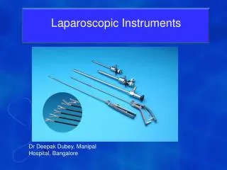
Surgical Approaches • Standard open approach • Laparoscopic (transanal removal specimen) • Laparoscopic-assisted (extraction incision only) • Hand-assisted laparoscopic • Hybrid Laparoscopic / Open method • TATA (Transanal – Transabdominal - Transanal)
Status of Laparoscopic TME & Rectal Resection for Cancer • Laparoscopic methods have been proven to be safe and effective for colon cancer • Far less data regarding rectal cancer resection • Randomized multi-center laparoscopic rectal cancer trials • COLOR 2 in Europe (over 850 patients entered) • ACOSOG Study (over 120 patients enrolled) • MITT Group (lap vs Hand LAR, just starting) • No long term prospective randomized results yet available • Single center data suggests lap TME possible
Rectal Resection For Cancer Only After Gaining Experience Doing Laparoscopic Colectomy • Should do rectal cases early only after: • Learning open TME methods • Learning 2 handed skills • Doing many lap colectomies • Do not attempt LAR early in your laparoscopic experience
Advantages of Laparoscopic Methods for TME • Superior visualization • Improved ability to identify: • Planes • Nerves • Vessels • Better able to do the distal portion of the mobilization sharply
Laparoscopic-Assisted LAR Resection: Port Placement Extraction site & possible stapling port
Laparoscopic Abdomino-Perineal Resection
Straight Laparoscopic LAR: The Start • Standard lateral to medial at left iliac fossa • Identify ureter & gonadal vessels • Mobilize main sigmoidal vessels • Enter posterior plane • Medial to lateral • Right side • Base of rectosigmoid • Near sacral promontory • Score parallel to the main sigmoidal vessels
Components of a TME (sphincter saving) • Posterior mesorectal mobilization • Lateral mesorectal mobilization • Anterior mesorectal mobilization • Distal mesorectal division • Distal rectal division • Anastomosis
Deep Pelvic Surgery • The bony pelvis limits outward traction • Important adjacent anterior structures • Bladder - Seminal vessicles • Prostate - Vagina • Important posterior structures • Hypogastric nerves - Nervi erigente • Presacral veins • Exposure is further limited in: • Males with narrow and long pelvis • Obese patients • Patients with large & bulky tumors
Retraction of the Giant Uterus • #2 nylon suture on straightened retention needle passed through lower abdominal wall • Once inside, needle passed through uterus near round ligament • Passed back outside • Tied over small gauze • Identical suture on opposite side
Other Methods of Uterine & Vaginal Retraction • Uterine manipulator • Retractor placed transvaginally into cervix • Fixed in position either with cervical balloon or a clamp • Downard traction on external end of device retracts the uterus upwards • Vaginal identification & retraction • Can use EEA sizers OR clean proctoscope
The Challenge of Transabdominal Closed Deep Pelvic Surgery Pubis Rectal transection level
Pubis Deep pelvis Rectum Side view Front view
Traction and Countertraction are Crucial ! The Assistant is the Key • Need 4 hands to do deep mobilization • Assistant provides much of the exposure • Choose dissection target • Posterior, anterior or lateral • Open atraumatic grasper is the tool • Apply strong traction & countertraction • Then retract cephalad !!! CRITICAL
Retract With Open Grasper Single point of retraction Two point retraction
Lateral Plane Exposure in Pelvis Colon & Rectum Bony confines of the pelvis Pubis
Exposing Left Lateral Plane Tissue Cutting Device Bowel graspers
Exposing Right Lateral Pelvis Tissue cutting device
Retraction to Expose R Side • Video clip 0002PowerPoint_Hi.wmv
Importance of Cephalad Retraction Element Video Clip: Gordon22Powerpoint_Hi.wmv:
Distal Rectal Retraction to Expose the Anterior Plane grasper Pubis Pelvis bladder Anterior peritoneal reflection 3 2 1 Rectum Leg Head Tucus Anus
Distal Rectal Retraction to Expose the Anterior Plane 2nd grasper Pubis Pelvis bladder Anterior peritoneal reflection 2 1 Rectum Leg Head Tucus Anus
Anterior Deep Dissection • In males: • Identify seminal vessicles • Leave Denonvillier’s fascia intact unless lesion is anterior • Avoid vas deferens (shouldn’t see it) • In females: • Find plane between vagina and anterior rectal wall • More fat in this space than you think
Extraperitoneal Rectal Mobilization • Alter traction until plane exposed • Shift dissection target frequently • Left lateral to anterior • Anterior to right lateral • Lateral to posterior • Pull back camera to get broader view • Find the clearest dissection field • When confused, change exposure and/or shift dissection target
Pelvic Tissue Division & Dissection in Open & Closed LAR • Monopolar cautery • Bipolar device • Ultrasonic shears • Avoid blunt dissection
Early Right Lateral Dissection • Video clip Cohen44PowerPoint_Hi.wmv
Laparoscopic-assisted Hand-assisted / Hybrid Full Open Incision Laparoscopic-assisted Full Open Incision Hand-assisted / Hybrid Full Open Incision Minimally Invasive Strategies
Hand and Hybrid Methods • Offer patients much of the benefits of MIS • Avoids full laparotomy • Do not have to fully complete case laparoscopically • Is a logical approach • If can take flexure down closed then patient will benefit.
Specimen Extraction Abdominal wall Extraction wound Specimen Abdominal cavity
Obesity Skin incision Fascial incision Peritoneal incision Skin incision Fascial incision Peritoneal incision