Download
1 / 23
500 likes | 1.65k Views
Laparoscopic Splenectomy. Jessica McQuerry University of Kentucky College of Medicine M1. Patient. Female in early 20’s presents with abdominal discomfort and feelings of early satiety On physical exam, a palpable mass is found in the left upper abdominal quadrant. Imaging.

E N D
Laparoscopic Splenectomy Jessica McQuerry University of Kentucky College of Medicine M1
Patient • Female in early 20’s presents with abdominal discomfort and feelings of early satiety • On physical exam, a palpable mass is found in the left upper abdominal quadrant
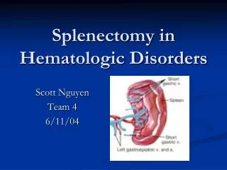
Imaging A CT scan was performed and showed a splenic cyst
Splenic Cysts • Uncommon • Incidence of 0.07% in general population • Majority of cases are due to parasitic infection with Echinococcusgranulosus resulting in hydatid disease • Non-parasitic cysts account for < 1/3 of all splenic cyst cases • Pseudo cyst (75%) • True cyst (25%)
treatment • Surgical cystectomy or splenectomy • Depends on the size of the splenic cyst • Depends on the position of the cyst in relation to the splenic hilum
Other indications • Indications: • Idiopathic thrombocytopenic purpura (ITP) • Autoimmune hemolytic anemia • Microspherocytosis • Benign tumors and cysts • AIDS-related thrombocytopenia • Relative contraindications: • Hematological malignancies • Moderate splenomegaly • Absolute contraindications: • Massive splenomegaly • Portal hypertension
Patient position • Right lateral decubitis, flexed at waist • A cushion is placed under the lumbar fossa to open up the operating field and facilitate trocar placement
Surgical team The surgeon faces the patient, the assistant is behind the patient. They each have their own video screen. The camera person stands next to the assistant.
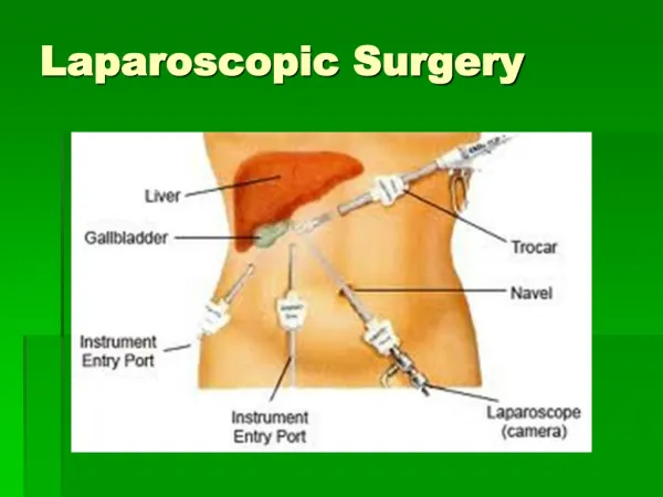
Trocar placements • Optical trocar, 10mm • Anterior axillary line below the left costal margin • Operating trocar, 5mm • Mid-axillary line below left costal margin • Operating trocar, 5mm • Mid-clavicular line, a few cm below the left costal margin • Retractor or operating trocar, 8-12mm • Mid-scapular line below the 12th rib
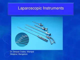
equipment • 30⁰ scope • Atraumatic graspers • Ultrasonic dissectors • Linear Stapler • L-hook Electrocautary tool • Flexible retractor • Suction-irrigation device • Specimen retrieval bag • Spleen scoop
procedure • Exploration • Check for mobility of the spleen and location of possible adhesions. 00:00- 6:40 • Exposure • Dissection of the splenophrenic ligament with the harmonic scalpel. 6:40- 8:43 & 18:30- 23:20
Procedure • Dissection of the splenocolic ligament. 9:14- 13:53 • Check for and remove any attachments to the abdominal wall. 13:53- 15:42
Procedure • Exposure and transection of the tissue and vessels in the gastrosplenic ligament. 23:20- 28:30 • An L hook cautery is used to dissect some of the retroperitoneal attachments. 28:30- 31:32
Procedure • Drainage of Cyst • Locate and drain the splenic cyst 32:40- 42:20
procedure Splenic Artery • Dissection of the splenorenal ligament. 42:20- 46:27 • Careful dissection of the splenic hilum. 46:27- 56:45 • Identify and staple the splenic artery. 56:45- 1:02:03 • Identify and staple the splenic vein. 1:02:03- 1:02:50
procedure • Detachment • Fully detach the spleen by removing any remaining attachments. 1:02:50- 0:40 (2) • Extraction • A bag is introduced in the retraction trocar. 3:30 (2)
Procedure • Insert the spleen in the bag and close. 3:30- 19:20 (2) • Pull the tip of the bag up through the retraction trocar. 31:20 (2) • The bag is cut away from the rim. • The spleen is morcellized with spleen scoops and removed.
Procedure • Closure • Check for tissue damage and accessory spleens 00:00- 5:31 (3)
Post operative care • Liquid diet- the night of or the morning after surgery • Regular diet and discharge from the hospital by the second postoperative day • Within two weeks, patients are usually able to return to work • Steroid dosages can be tapered rapidly and then discontinued
complications • Intraoperative complications: • Uncontrollable bleeding • Injury to regional organs during dissection • More common with larger spleens • Postoperative complications: • Minor wound infections • Postoperative ileus • Infection • ITP: • Recurrent or persistent decrease in the number of blood platelets • Chronic ITP
outcomes • Curative in about 50-60 percent of patients • Improves another 20-35 percent • Fails to help 5-10 percent
Laparoscopic vs. open • Primary benefit of laparoscopic is several small incisions instead of one large incision • Shorter hospital stay • Quicker recovery • Better cosmetic result • Laparoscopic procedure is a more demanding technique • Highly vascularized organ • Fragile parenchyma • Attached by several ligaments to other organs • Hematological disease often associated with a low platelet count
references • Targarona, EM. (2002, March). Laparoscopic splenectomy: anterior posterior approach. Retrieved from http://www.websurg.com/ref/media.php?doi=ot02en199a • The University of Texas Southwestern Medical Center at Dallas. (2010). Laparoscopic spleen surgery. Retrieved from http://www8.utsouthwestern.edu/utsw/cda/dept48035/files/89885.html • Adas, G, et al. (2009). Diagnostic problems with parasitic and non-parasitic splenic cysts. BMC Surgery , 9(9), Retrieved from http://www.biomedcentral.com/1471-2482/9/9doi: 10.1186/1471-2482-9-9 • KalinovaK. (2005). Giant pseudocyst of the spleen: A case report and review of the literature. Journal of Indian Association of Pediatric Surgeons, 10(3), Retrieved from http://www.bioline.org.br/request?ip05044