Download
1 / 51
1.02k likes | 2.36k Views
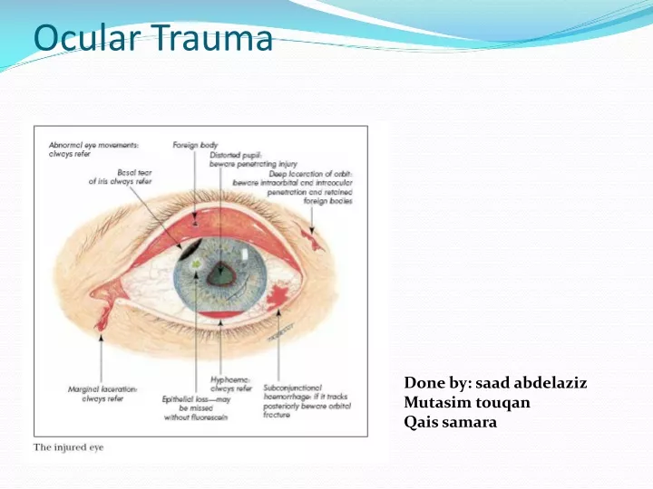
Ocular Trauma. Done by: saad abdelaziz Mutasim touqan Qais samara. Learning Objectives. To take a good history in a case of ocular trauma To understand the effects of trauma on the eye To understand the management of eye trauma To understand basic terms related to eye trauma.

E N D
Ocular Trauma Done by: saad abdelaziz Mutasim touqan Qais samara
Learning Objectives • To take a good history in a case of ocular trauma • To understand the effects of trauma on the eye • To understand the management of eye trauma • To understand basic terms related to eye trauma
Introduction Although the eye is well protected by the orbit, it may yet be subject to injuries. forms of injury include: • Foreign bodies • Blunt trauma • Penetrating trauma • Chemical and radiation injuries
risk factors • Gender : 75%-80% of them are in males • Age: more in children and young age group • Occupation : construction, industry • Sports : boxing , racket sports • Motor vehicle accidents
Effects of eye injury : • Closed globe injury or Non-penetrating trauma: The eye globe is intact, but the seven rings of the eye have been classically described as affected by blunt trauma. • Penetrating trauma: The globe integrity is disrupted by a full-thickness entry wound and may be associated with prolapse of the internal contents of the eye.
Effects of eye injury (cont.) • Blowout fracture of the orbit is caused by blunt trauma, classically described for fist or ball injury, leading to fracture of the floor or medial wall of the orbit due to sudden increased pressure on the orbital contents. • Perforating trauma: The globe integrity is disrupted in two places due to an entrance and exit wound (through and through injury). This is a quite severe type of eye injury.
Foreign bodies Corneal foreign body is foreign material on or in the cornea, usually metal, glass, or organic material.
Corneal foreign bodies: Symptoms Foreign body sensation, Tearing, History of trauma ,photophobia , pain , red eye Signs Corneal foreign body with or without rust ring, edema of the lids, conjunctiva, and cornea, foreign body can cause infection and/or tissue necrosis.
Corneal foreign bodies cont. Workup 1.History 2.Document visual acuity. One or two drops of topical anesthetic may be necessary to control pain. 3.Slit-lamp Examination: If there is no evidence of perforation, evert the eyelids and inspect for foreign bodies. 4.Dilate the eye and examine the vitreous and retina 5.Consider a B-scan US, CT of the orbit.
Corneal foreign bodies cont. Treatment 1.Apply topical anesthetic, remove the foreign body with a spud or forceps at a slit lamp. If multiple superficial foreign bodies, its easier to remove with irrigation. 2.Remove the rust ring. This may require an ophthalmic drill. 3.Measure the size of the resultant corneal epithelial defect. 4.Treat as for corneal abrasion.
Blunt trauma blunt impact may damage the structures at the front of the eye (the eyelid, conjunctiva, sclera, cornea, iris, and lens) and those at the back of the eye (retina and optic nerve). If a small objects ( such as a squash ball, shuttlecocks, knuckles, etc.) hits the area the eye itself may take most of the impact. If a large object (such as a football, or by fist) hits the eye most of the impact is usually taken by the orbital margin. Such an impact may also result in damage to the orbit (blow-out fracture).
Penetrating trauma when a foreign body passes through the ocular coat of the eye, this will cause damage in the ocular structures, and in some cases the foreign body may also be retained in the eye. penetrating injury of the eye represents a major threat to vision in the workplace,home and school.
Lid lacerations: Eyelid Lacerations: Cuts to the eyelid caused by trauma Superficial Lacerations can be usually treated in the emergency room under local anesthesia
Subconjunctival hemorrhage: Is bleeding underneath the conjunctiva. The conjunctiva contains many small, fragile blood vessels that are easily ruptured or broken. When this happens, blood leaks into the space between the conjunctiva and sclera. Symptoms Red eye, may have mild irritation, usually asymptomatic Signs Blood underneath the conjunctiva, often in a sector of the eye. The entire view of the sclera may be obstructed by blood. Causes Valsalva (e.g., coughing or straining), Trauma, HTN, Bleeding disorder, Hemorrhage due to orbital mass (rare), Idiopathic.
Subconjunctival hemorrhage cont. Workup -History: Bleeding or clotting problems? Medications (e.g., aspirin, warfarin)? Eye rubbing, trauma, heavy lifting, Valsalva? Recurrent Subconjunctival Hemorrhage? Acute or Chronic cough (COPD)? -Check Vital signs -History of recurrence or bleeding problem; order Bleeding time, PT, PTT, CBC. -Positive Orbital signs: CT scan with and without contrast
Subconjunctival hemorrhage cont. -Ocular Examination: Rule out a conjunctival lesions, Check IOP, and Check extraocular motility. In traumatic cases you should rule out:Ruptured Globe (Abnormal deep ant. Chamber, Significant SCH, Hyphema, Vitreous hemorrhage, or prolapse of uveal tissue) . Retrobulbar Hemorrhage (Exophthalmus, Increased IOP, and chemosis) Orbital Fracture (Limited extraocular eye motility, eno- or exo-phthalmus, preiorbital crepitus, paraesthesia.
The cornea • The cornea is the transparent front part of the eye that covers the iris, pupil, and anterior chamber. Together with the lens, the cornea refracts light, accounting for approximately two-thirds of the eye's total optical power.[ • Corneal epithelium: a thin epithelial multicellular tissue layer (non-keratinized stratified squamous epithelium • Bowman’s layer • Corneal stroma • Descemet’s membrane • Corneal endothelium
Corneal abrasion: is a medical condition involving the loss of the surface epithelial layer of the eye’s cornea. It’s the most common eye injury and perhaps one of the most neglected , it occurs because of a disruption of the integrity of corneal epithelium because the corneal surface scraped away or denuded as a result of physical external forces.
Corneal abrasion cont. • They usually heal without serious consequences, although deep abrasion can result in scar formation in the stroma. • Examples about causes of corneal abrasion : corneal or epithelial disease (eg, dry eye), superficial corneal injury or ocular injuries (eg, those d.ue to foreign bodies), and contact lens wear .
Corneal abrasion cont. • Most patients present with the following: Photophobia Watering Foreign body sensation Gritty feeling Pain • Signs: Corneal edema Bacterial corneal ulcers Fungal, amebic, or viral corneal ulcers Uveitis
Patients may be troubled by recurrent episodes of pain particulary in the early hours of the morning or on waking. • This condition is termed “recurrent corneal abrasion” • It happens due to adhesion of the resurfacing epithelium to bowman’s layer at the site of injury • Prophylaxis against recurrent corneal erosion can be achieved by using lubricating ointment at night.
Corneal abrasions cont. Treatment: • Antibiotics Ointment (Erythromycin, Ciprofloxacin) Drops (Polytrim, Fluoroqunilone) • Cycloplegic agent(Cyclopentolate) for discomfort from traumatic iritis which may develop 24-72 hours. AVOID STEROIDS. • Patching for comfort, and avoiding scratching of the eye during sleep. • Topical NSAIDS drops (Ketorolac) for pain control. AVOID in post-op patients • Debriding loose or hanging epithelium • No contact lens wear
Corneal lacerations: A corneal laceration is a partial- or full-thickness injury to the cornea. A partial-thickness injury does not violate the globe of the eye (abrasion). A full-thickness injury penetrates completely through the cornea, causing a ruptured globe
Corneal laceration Partial thickness: Signs The Ant. Chamber isn’t entered, therefore, the cornea isn’t perforated Workup 1.Complete ocular examination 2.Seidel test. If positive then it’s a full-thickness laceration. Seidle test: is used to assess the presence of anterior chamber leakage in the cornea.
Corneal lacerations cont. Treatment 1. Cycloplegic (Scopolamine) and an antibiotic (Polysporin, Fluroquinolone drops) 2.If moderate to deep corneal laceration is accompanied by wound gape, it is often best to suture. 3.Tetanus toxoid for dirty wounds Follow up Reevaluate daily until the epithelium heals.
Corneal lacerations cont. Full thickness: We should exclude Ruptured Globe and Penetrating Ocular injury, A full-thickness injury will allow aqueous humor to escape the anterior chamber, which can result in a flat-appearing cornea, air bubbles under the cornea, or an asymmetric pupil secondary to the iris protruding through the corneal defect. Small, self-sealing, or slow leaking lacerations may be treated with aqueous suppressants, bandage soft contact lenses, fluroquinolone drops. Alternatively, a pressure patch and twice-daily antibiotics may be used. AVOID steroids.
Hyphema: Blood in the Anterior Chamber Symptoms Pain, Blurred vision, History of blunt trauma Signs Blood in the Anterior Chamber. Gross layering or clot or both, usually visible without a slit lamp. A total (100%) hyphema may be black or red; when black its called “8-ball” or “black ball” hyphema.
Hyphema cont. Workup 1. History: Mechanism of injury, approximate time and day, time of visual loss, Medications (Aspirin, NSAIDs, Warfarin), History or family history of sickle cell disease/traits. 2. Complete Ocular Examination 3. CT scan of the orbit 4. Screen for sickle cell disease or trait Factors with poor outcome: 1. Poor visual acuity (worse than 20/200) 2. Sickle cell disease/trait with increased IOP 3. Medically uncontrollable IOP 4. Large initial hyphema 5. Recent Aspirin, NSAIDs use 6. Delayed presentation
Hyphema cont. Treatment For all patients 1. Complete bed rest or hospitalization 2. Place a shield over the injured eye . Elevation of the head of the bed by approximately 45 degrees (so that the hyphema can settle out inferiorly and avoid obstruction of vision, as well as to facilitate resolution 3. Atropine 4. Mild analgesics 5. Topical steroids drops (Traumatic iritis develop 2-3 days) 6. NO aspirin or NSAIDs .
Causes:hyphaemas are frequently caused by injury • “blunt truma”,and it may partially or completely block vision. • complications:1.hemosiderosis • 2.hetrochromia • 3.blood acumulation may also cause elevtion of the intraocular pressure
of the lens:Subluxation /dislocation Definition: Subluxation: Partial disruption of the zonular fibers; the lens is decentered but remains partially in the pupillary aperture Dislocation: Complete disruption of the zonular fibers; the lens is displaced out of the papillary aperture.
Subluxation/dislocation of the lens cont. Symptoms Decreased vision, double vision that persists when covering one eye (monocular diplopia) Sign Decentered or displaced lens,. Marked astigmatism, Cataract, Angle-closure glaucoma as a result of pupillary block, acquired high myopia, viterous in the ant. Chamber, asymmetry of the ant. Chamber depth Causes 1. Trauma most common cause 2. Marfan Syndrome 3. Homocystinuria
Subluxation/dislocation of the lens cont. Treatment 1. Subluxation: Asymptomatic; Observe High uncorrectable astigmatism; Surgical removal of the lens Symptomatic cataract: Surgical removal, Mydriasis (Scopolamine), pupillary constriction (Pilocarpine), and phakic correction. 2. Pupillary block 3. In dislocation; surgical intervene
:Traumatic cataract Traumatic cataracts occur secondary to blunt or penetrating ocular trauma. Infrared energy , and ionizing radiation are other rare causes of traumatic cataracts. Cataracts caused by blunt trauma classically form stellate- or rosette-shaped. penetrating trauma with disruption of lens capsule forms cortical changes
Traumatic cataract cont. History Mechanism of injury - Sharp versus blunt Past ocular history - Previous eye surgery, glaucoma, retinal detachment, diabetic eye disease Planning surgical approach is of most importance in cases of traumatic cataract.
Blowout fracture A blowout fracture is a fracture of the walls or floor of the orbit. Intraorbital material may be pushed out into one of the paranasal sinuses. This is most commonly caused by blunt trauma of the head
:Blowout fracture • Symptoms: Pain (especially on attempted vertical eye movement) Local tenderness Binocular double vision Eyelid swelling And creptius after nasal blowing • Sign: damage to the orbit itself is suspected if the following signs are present : • Emphysema (air under the skin with crackles when pressed) derived from the fractured sinus. • Parasthesia below the orbital rim suggesting infraorbital nerve damage • Limitation of eye movement , particularly on upgaze and downgaze , due to tethering of the inferior rectus muscle .
Blowout fracture cont. -Treatment(most adult orbital fractures can initially be followed conservatively) *Broad spectrum oral antibiotic (may be use but not mandatory) *Instruct the patient not to blow his nose *Apply ice packs to the orbit for the first 24 to 48 hours The aim of treatment is prevention of permanent diplopia and cosmetically unacceptable enophthalmos. The factors that determine the risk of late complications are -Fracture size -Herniation of orbital content into the maxillary sinus -Muscle entrapment *Surgical repair -Immediate repair (usually within 24hr.) -Repair in 1 to 2 weeks *Neurosurgical consultation is recommended
Restriction on upgaze due to trapping of the inferior rectus muscle by connective tissue septa caught in the fractured site. The inferior orbital floor is the most commonly fractured site.
Commotio retinae: Concussion of the retina that may produce a milky edema in the posterior pole that clears up after a few days. Symptoms Decreased vision or asymptomatic, history of recent ocular trauma Signs Confluent area of retinal whitening
Commotio retinae cont. Workup Complete ophthalmic examination, including dilated fundus examination. Scleral depression is performed excep when a hyphema, or iritis is present Treatment No treatment is required because this condition usually clears without therapy Follow up Dilated fundus examination is repeated in 1-2 weeks.
Traumatic retinal detachment: Retinal detachment refers to separation of the inner layers of the retina from the underlying retinal pigment epithelium (RPE, choroid). Emergency Department treatment of retinal detachment consists of evaluating the patient and treating any unstable vital signs, preparing the patient for possible emergency surgery.
Chemical burn (injury) • Most chemical substances that come in contact with the conjunctiva or cornea cause little harm. • The chief danger comes from alkali-containing compounds found in household cleaning fluids, fertilizers and pesticides. They erode and opacify the cornea. • Acid-containing compounds (battery fluid, chemistry labs) are somewhat less dangerous. • There are no antidotes to these chemicals. The best you can do is to dilute them immediately with plain water. • The resultant reaction of the tissue causes the damage.
Chemical burn (injury) cont. Treatment should be instituted immediately, even before testing vision. Emergency treatment: 1-copious irrigation of the eyes, preferably with saline or ringer lactate. Don’t use acidic solutions to neutralize alkalis or vice versa. Pull down the lower eyelid and evert the upper eyelid to irrigate the fornices 2-irrigation should be continued until neutral PH is reached. The volume of irrigation fluid required to reach neutral PH varies with the chemical and the duration of the chemical exposure
Chemical burn (injury) cont. For mild to moderate burns (during and after irrigation): • cycloplegic • topical antibiotic • oral pain medication • if increase IOP use drugs to reduce it (acetazolamide, methazolamide add b blocker if additional IOP control is required) • frequent use of preservative free artificial tear
Chemical burn (injury) cont. For severe burns (Treatment after irrigation): • Admission to the hospital Lysis of conjunctival adhesion • Debride necrotic tissue • Topical antibiotic • Topical steroid • Consider a pressure patch • Antiglaucoma medication if the IOP is increased or cant be determined • Frequent use of preservative free artificial tear • Other consideration: Therapeutic contact lenses, collagen, amniotic membrane transplant IV ascorbate and citrate for alkali burns If any melting of the cornea occurs, collagenase inhibitors may be used If the melting progresses an emergency patch graft or corneal transplat may be necessary.
Chemical burn (injury) A hazy cornea following an alkali burn