Download
1 / 46
460 likes | 496 Views
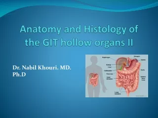
Anatomy and Histology of the GIT hollow organs I. Dr. Nabil Khouri . MD. Ph.D. Digestive System Anatomy. Digestive tract Alimentary tract or canal Gastrointestinal (GI) tract Accessory organs Primarily glands Liver, gallbladder, pancreas, salivary glands Regions Mouth or oral cavity

E N D
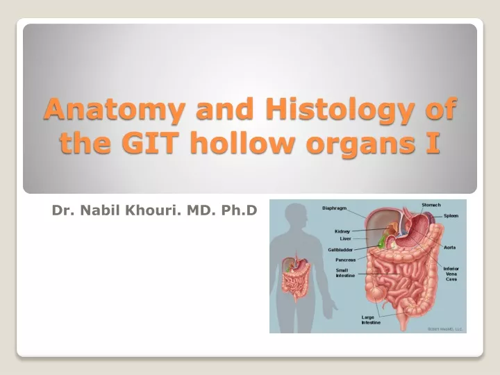
Anatomy and Histology of the GIT hollow organs I Dr. NabilKhouri. MD. Ph.D
Digestive System Anatomy • Digestive tract • Alimentary tract or canal • Gastrointestinal (GI) tract • Accessory organs • Primarily glands • Liver, gallbladder, pancreas, salivary glands • Regions • Mouth or oral cavity • Pharynx • Esophagus • Stomach • Small intestine • Large intestine • Anus
Is a muscular bag-most distensible part of the digestive tract. Occupies the pigastric, umbilical & left hypochondrium regions. It is a J-shaped to pyriform structure. The exact size, shape and position of the stomach can vary from person to person. For example, in thin individuals, it is not uncommon for the stomach to extend into the pelvic region.. Anatomical position of the Stomach
EXTERNAL FEATURES • Two orifices : Cardiac and Pyloric • Two curvatures : Greater and Lesser • Two surfaces : Anterosuperior and Posteroinferior
Stomach Anatomy Fundus Esophagus Longi- tudinal layer (outer) Cardia Pyloric orifice Pyloric sphincter Three layers of smooth muscle Circular layer (middle) Duodenum Oblique layer (inner) Pylorus Body Gastric folds • Openings • Gastroesophageal: to esophagus • Pyloric: to duodenum
Sphincters of the Stomach • The inferior Esophageal sphincter is a (physiological) located between the oesophagus and the stomach . • It is located to the left of the T11 vertebra. Situated immediately superior is the Esophageal hiatus. • Histologically, the sphincter is marked by an abrupt change from stratified squamous epithelium to simple columnar. • At the Esophageal hiatus, the right crus of the diaphragm exerts pressure on the Esophageal wall. • In the normal (non-eating) state, the lumen of the Esophagus is collapsed. • The Esophagus enters the stomach at an angle – when the stomach is full, this closes off entry to the stomach.
Pyloric Sphincter • The pyloric sphincter lies between the pylorus and the duodenum. • It controls of the exit of chyme (food and gastric acid mixture) from the stomach. • In contrast to the inferior Esophageal sphincter, this is an Anatomical sphincter. It contains smooth muscle, which constricts to limit the discharge of stomach contents through the orifice. • The pylorus is normally contracted so that the orifice is small and food can stay in the stomach for a suitable period.
Parts of the stomach Greater curvature – forms the long, convex, lateral border of the stomach. Arising at the cardiac orifice, it arches backwards and passes inferiorly to the left. It curves to the right as it continues medially to reach the pyloric antrum. The short gastric arteries and the right and left gastro-omental arteries supply branches to the greater curvature. Lesser curvature – forms the shorter, concave, medial surface of the stomach. The most inferior part of the lesser curvature, the angular notch, indicates the junction of the body and pyloric region. The lesser curvature gives attachment to the hepatogastric ligamentand is supplied by the left gastric artery and right gastric branch of the hepatic artery.
Cardiac region (cardia) muscularis externa (longitudinal, circular, oblique muscles) • Body • Pyloric region (pyloric antrum, pyloric canal, pylorus, pyloric sphincter valve) • Lesser momentum (liver to lesser curvature) • Greater momentum (greater curvature to cover small intestine)
STOMACH RELATIONS • Peritoneal Relations
VENOUS DRAINAGE • Right & Left gastric veins-Portal vein • Right gastroepiploic vein-superior mesenteric vein • Left gastroepiploic & Short gastric veins-Splenic vein
Microscopic Anatomy Stomach Histology • The four tunics typical of the alimentary canal • Mucosa • Submucosa • Muscularis Externia • Serosa
Rugae: Folds in stomach when empty • Gastric pits: Mark the Openings for gastric glands • Common secreting cells • Mucous cells: Mucus along surface and in pits • Parietal cells: Hydrochloric acid • Chief cells: Pepsinogen
Mucus neck cells produce a different type of mucus of the surface epithelium • The special function of this unique mucus is not yet understood • Chief cells produce pepsinogen, the inactive form of the protein- digesting enzyme pepsin • Parietal cells scattered among the chief cells secrete hydrochloric acid (HCl) and intrinsic factor • The parietal cells have a large surface area adapted for secreting HCl in the stomach
Microscopic Anatomy • Enteroendocrine cells found in the gastric glands secrete various hormones into the interstitial fluid of the lamina propria. These include gastrin, which is released mainly by enteroendocrine G • Intrinsic factor is required for absorption of B12 in the small intestine
The Small Intestine • About 18 feet long • 3 regions • The duodenum • Common bile duct & pancreatic duct empty here • The jejunum • The ileum • Ends in the ileocecal valve
First part of duodenum • It begins at the pylorus and runs upward and backward on the transpyloric plane at the level of the 1st lumbar vertebra • The relations of this part are as follows: • ■■ Anteriorly: The quadrate lobe of the liver and the gallbladder • ■■ Posteriorly: The lesser sac (first inch only), the gastroduodenal artery, the bile duct and the portal vein, and the inferior vena cava • ■■ Superiorly: The entrance into the lesser sac (the epiploic foramen) • ■■ Inferiorly: The head of the pancreas
runs vertically downward in front of the hilum of the right kidney on the right side of the 2nd and 3rd lumbar vertebrae. About halfway down its medial border, the bile duct and the main pancreatic duct pierce the duodenal wall. They unite to form the Ampulla that opens on the summit of the major duodenal papilla. The accessory pancreatic duct, if present, opens into the duodenum a little higher up on the minor duodenal papilla. The second part of the duodenum
Relation of the second part of duodemum • ■■ Anteriorly: The fundus of the gallbladder and the right lobe of the liver, the transverse colon, and the coils of the small intestine • ■■ Posteriorly: The hilum of the right kidney and the right ureter • ■■ Laterally: The ascending colon, the right colic flexure, and the right lobe of the liver • ■■ Medially: The head of the pancreas, the bile duct, and the main pancreatic duct
The third part of the duodenum • it runs horizontally to the left on the subcostal plane, passing in front of the vertebral column and following the lower margin of the head of the pancreas • Relations of this part are : • ■■ Anteriorly: The root of the mesentery of the small intestine, the superior mesenteric vessels contained within it, and coils of jejunum • ■■ Posteriorly: The right ureter, the right psoas muscle, the inferior vena cava, and the aorta • ■■ Superiorly: The head of the pancreas • ■■ Inferiorly: Coils of jejunum Fourth Part of the Duodenum
The fourth part of the duodenum • it runs upward and to the left to the duodenojejunal flexure .The flexure is held in position by a peritoneal fold, the ligament of Treitz, which is attached to the right crus of the diaphragm. The relations of this part are as follows: • ■■ Anteriorly: The beginning of the root of the mesentery and coils of jejunum • ■■ Posteriorly: The left margin of the aorta and the medial border of the left psoas muscle
Histology of the small intestine key points • The outermost layer of the intestine, the serosa, is a smooth membrane consisting of a thin layer of cells that secrete serous fluid, and a thin layer of connective tissue. • The muscularis is a region of muscle adjacent to the submucosa membrane. It is responsible for peristalsis movement . It usually has two distinct layers of smooth muscle: circular and longitudinal. • The submucosa is the layer of Dense Irregular CT or loose CT that supports the mucosa • The mucosa is the innermost tissue layer of the small intestines and is a mucous membrane that secretes digestive enzymes and hormones. The intestinal villi are part of the mucosa. • The three sections of the small intestine look similar to each other at a microscopic level, but there are some important differences. The jejunum and ileum do not have Brunner's glands in the submucosa, while the ileum has Peyer's patches in the mucosa, but the duodenum and jejunum do not.
Villi:● Short and stubby in duodenum, very tall in jejunum, intermediate height in ileum● Contain microvilli● Villus to crypt length is 3-5:1● Lined by primarily columnar absorptive cells and goblet cells● Villi may be short and distorted next to lymphoid aggregates● Each villus contains an arteriole with capillary network, veins and a central lymphatic with numerous nerve fibres
The wall of the small intestine is composed of four layers, from the outside to the inside: serosa, muscularis, submucosa, and mucosa. Brunner's glands Compound, tubular, submucosal glands found in that portion of the duodenum that is above The hepatopancreatic sphincter (sphincter of Oddi). The main function of these glands is to produce a mucus-rich, alkaline secretion (containing bicarbonate) in order to neutralize the acidic content of chyme that is introduced into the duodenum from the stomach, and to provide an alkaline condition for optimal intestinal enzyme activity Peyer's patches Patches of lymphoid tissue or lymphoid nodules on the walls of the ileum in the small intestine.
https://legacy.owensboro.kctcs.edu/gcaplan/anat2/histology/histo%20J%20digestive%20system.htmhttps://legacy.owensboro.kctcs.edu/gcaplan/anat2/histology/histo%20J%20digestive%20system.htm Crypts of Lieberkuhn:● Lower 20% of epithelium, contain undifferentiated (immature) crypt cells, Paneth cells (have large, apical eosinophilic granules containing antimicrobial proteins), scattered goblet cells and endocrine cells● Are surrounded by pericrypt fibroblast sheath● Secrete ions, water, IgA, antimicrobial peptides into lumen● Crypt cells take 3-8 days to migrate to surface● Allows for rapid repair, but also causes these cells to be sensitive to radiation therapy and chemotherapy