Download
1 / 14
160 likes | 418 Views
Histology for Pathology Lymphoid Organs. Theresa Kristopaitis , MD Associate Professor Director of Mechanisms of Human Disease Kelli A. Hutchens, MD, FCAP Assistant Professor Assistant Director of Mechanisms of Human Disease Loyola Stritch School of Medicine.

E N D
Histology for PathologyLymphoid Organs Theresa Kristopaitis, MD Associate Professor Director of Mechanisms of Human Disease Kelli A. Hutchens, MD, FCAP Assistant Professor Assistant Director of Mechanisms of Human Disease Loyola Stritch School of Medicine
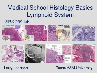
Lymphoid OrgansLymph Nodes, Spleen, Thymus • List the main function(s) of lymph nodes. • On an H&E stained section of a lymph node identify the capsule, subcapsular sinus, cortex, and medulla. • Identify the germinal center of a secondary lymphoid follicle. • Outline the path of lymph through a lymph node. • On an H&E stained slide of the spleen, identify the capsule, white pulp (central artery, nodule) and red pulp. • Summarize the key functions of the white pulp and red pulp of the spleen. • On an H&E stained section of an infant/child thymus identify the cortex and medulla of thymic lobules. • Define “Hassal corpuscle” and identify on a histologic section. • Summarize the key function(s) of the thymus. • Define “Mucosa-Associated Lymphatic Tissues” and list in which organs they are found.
Lymph Node Cortex Capsule MEDULLA
Lymph node Subcapsular Sinus
Lymph Node Follicle with Germinal Center (Secondary Follicle)
Pale medullary sinuses surrounding darker medullary cords. You can see many stellate reticular cells which, with reticular fibers, make a meshwork through the sinuses. Lymph "percolates" through the meshes of the sinuses while debris of foreign matter in it is phagocytized.
Spleen Red pulp White pulp
Spleen Red pulp White pulp
White Pulp Lymphoid nodule Central artery
Spleen Red Pulp Red pulp consists of open sinuses and cellular cords
ThymusInfant cortex Medulla
Thymus Medulla Hassall’s Corpuscle
Adult Thymus Panoramic view of adult thymus, largely replaced with adipose tissue. There are recognizable remnants of thymic lymphatic tissue, however, and Hassall's corpuscles are still present in the medulla.