Download
1 / 61
700 likes | 1.28k Views
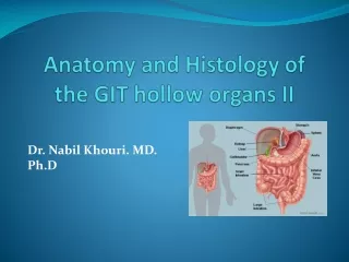
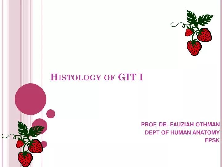
Histology of GIT I. PROF. DR. FAUZIAH OTHMAN DEPT OF HUMAN ANATOMY FPSK. Content. Histology of the: - oral cavity - pharynx - peritoneum - oesophagus - stomach - small intestine - large intestine. Digestive System. Digestive System. Structures involved in digestive system.

E N D
Histology of GIT I PROF. DR. FAUZIAH OTHMAN DEPT OF HUMAN ANATOMY FPSK
Content Histology of the: - oral cavity - pharynx - peritoneum - oesophagus - stomach - small intestine - large intestine
Structures involved in digestive system The digestive system of mammals consists of the following: -(mouth, oral cavity, pharynx, esophagus, stomach, small intestine, large intestine, rectum, anus) -also includes other associated structures/organs/glands (salivary glands, gall bladder, liver, pancreas).
Function of Digestive System Digestion is the process of breaking down food into molecules small enough for the body to absorb.
Oral Cavity Diagram uvula
Oral Cavity • Salivary glands produce large amounts of saliva • Saliva contains: • water for moistening food • Mucus for lubricating food and binding it into a bolus (ball of mush) • salivary amylase to start the breakdown of starch
Oral cavity • Oral or buccal cavity: • Is bounded by lips, cheeks, palate, and tongue • To withstand abrasions: • The mouth is lined with stratified squamous epithelium • The gums, hard palate, and dorsum of the tongue are slightly keratinized
How is food swallowed? Food moves to the pharynx, (throat) which in humans, leads to both the trachea and the esophagus. While food is being swallowed, the epiglottis blocks the trachea and the uvula blocks off the nose. Then food reaches the esophagus,(tube that connects the pharynx to the stomach)
(throat) uvula
Pharynx • Lined with stratified squamous epithelium and mucus glands • Has two skeletal muscle layers • Inner longitudinal • Outer pharyngeal constrictors
Peritoneum • Peritoneum – serous membrane of the abdominal cavity • Visceral – covers external surface of most digestive organs • Parietal – lines the body wall • Simple squamous epithelium
Oesophagus • Esophageal mucosa – nonkeratinized stratified squamous epithelium • The empty esophagus is folded longitudinally and flattens when food is present • Glands secrete mucus as a bolus moves through the esophagus • Muscularis changes from skeletal (superiorly) to smooth muscle (inferiorly)
General histology of GIT • Consist of 4 layer arrangement of tissue. • Mucosa • Submucosa • Muscularis • Serosa
Stomach The stomach has several muscle layers surround the stomach, serving to churn food. The stomach can expand to hold about 2 L of food (= 1/2 gal). It contains acid to digest food (ph = 2) and enzymes to breakdown protein.
Sphincters The cardiac sphincter closes off the top end of the stomach and the pyloric sphincter closes off the bottom
Stomach The stomach has three layers of muscle: • an outer longitudinal layer, • a middle circular layer, • an inner oblique layer. The inner lining consists of four layers: • the serosa, • the muscularis, • the submucosa, and • the mucosa.
The mucosa is densely packed with gastric glands, which contain cells that produce digestive enzymes, hydrochloric acid, and mucus.
Small Intestine and accessory organs Small intestine A lot of digestion happens here. S. int. secretes enzymes and pancreas/gall bladder dump enzymes into duodenum to continue digestion. Liver- The largest internal organ of the body. Makes bile, which aids in the digestion of fat. Detoxifies poisons like alcohol. Stores extra glucose in the form of glycogen. Gall Bladder- Sack on the bottom of one of the liver lobes. Stores bile until it is ready to move into the duodenum. Pancreas- Makes digestive enzymes that are dumped into duodenum of the small intestine. Makes the hormone, insulin, which regulates the amount of sugar in the blood.
Small Intestine SECTIONS OF THE SMALL INSTESTINE Duodenum- The first part of the small intestine which has ducts (tubes) leading into it from the liver/gall bladder and pancreas. Bile and pancreatic enzymes are mixed with food here. Jejuno-ileum- All the small intestine except for the duodenum. Digestion of food is completed here and nutrients are absorbed through its walls into the blood stream. Caecum-a pouch off the digestive tract between the small intestine and the colon. Produces enzymes that digest cellulose. (is the appendix in humans)
Small intestine • Small intestine, which has three parts: • Duodenum • Jejunum • Ileum
The epithelial component of the small intestine is composed of villi (finger like projections) and crypts (crypts of Liberkuhn).
Large intestine • Large intestine, which has three parts: • Cecum (the vermiform appendix is attached to the cecum). • Colon (ascending colon, transverse colon, descending colon and sigmoid flexure) • Rectum Plicae circularis (valves of Kerckring) – transverse semilunar folds that contain a core of submucosa
Large Intestine The large intestine or colon functions to re-absorb water. Bacteria live here like Escherichia coli (E. coli) which produce gases as they ferment their food. Occasionally, some of this gas is released. As these bacteria digest/ferment left-over food, they secrete beneficial chemicals such as vitamin K, Vitamin B, and some amino acids, and are our main source of some of these nutrients.
Sections of Large Intestine Spiral colon-spiraled part of the large intestine. Absorbs water, vitamins, and minerals from the food and moves them into the bloodstream. Descending colon- The part of the large intestine leading from the spiral colon down to the rectum. Same function as the spiral colon.
Structure of LI; • simple columnar epith • Crypts of Leiberkuhn • lymph tissue (GALT) • goblet cells • NO kerckring folds or villi or paneth cells • Layers: Mucosa, Submucosa, Musc. Ext, and either serosa & adventia