Download
1 / 33
340 likes | 477 Views
HYPOXIA. Dr B.Kalpana. At the end of class students should be able to: 1.Define and classify hypoxia 2. Explain the causes and characteristics features of each type of hypoxia. 3.What is oxygen therapy and its uses in hypoxia.

E N D
HYPOXIA Dr B.Kalpana
At the end of class students should be able to: 1.Define and classify hypoxia 2. Explain the causes and characteristics features of each type of hypoxia. 3.What is oxygen therapy and its uses in hypoxia. 4. Define cyanosis. Mention its types and its sites of appearance on the body.
O2 deficiency occurs at tissue level due to -- either due to Decrease O2 content in the blood –which may be due to (1) Decrease O2 tension in arterial blood (2) Decrease O2 carrying capacity of blood Or Decrease O2 supply by decrease blood flow Or Decrease O2 utilization by the tissue
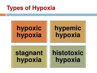
Classification of hypoxia • Hypoxic hypoxia or Arterial hypoxia • Anemic hypoxia • Stagnant hypoxia or Ischemic hypoxia or Hypo kinetic hypoxia • Histotoxic hypoxia
Hypoxic hypoxia • Inadequate PO2 in arterial blood (PaO2). May results from: • Inadequate PO2 in inspired air • Major hypoventilation • Inadequate alveolar – capillary transfer-defect in exchange of gases-affecting V/P ratio. • Venous –arterial shunts.-congenital heart disease.
Hypoxic Hypoxia Reduced pO2 in the lungs (high altitude) Red blood cells Body tissue
Partial pressure of oxygen –decreases • 0xygen carrying capacity –normal • Oxygen content in blood-decreases • Blood flow rate and tissue utilization –normal • A-V of partial pressure ofoxygen decreases • Cyanosis is present
Anaemic Hypoxia • PaO2 normal but concentration of functional haemoglobin is reduced. • Possible causes of anaemia: • Deficiencies of iron, vitamin B12, folate or copper • Kidney disease affecting production of erythropoietin • Excessive blood loss • Carbon monoxide poisoning
CO poisoning • CO has 200 times more affinity to Hb than O2. • CO with Hb is Carboxyhemoglobin (HbCO) • HbCO is cherry red color • HbCO cannot take O2 & liberation of CO from Hb also is slow • Also HbCO shifts remaining HbO2 shift to left • Hyperventilation is absent as arterial PO2 is normal—no peripheral chemoreceptor drive • Symptoms –like other hypoxia –headache, nausea etc. • Hyperbaric O2 therapy is useful
Partial pressure of oxygen –normal • 0xygen carrying capacity –decreases • Oxygen content in blood-decreases(moderately) • Blood flow rate and tissue utilization –normal • A-V 0f Partial pressure of oxygen -normal • Cyanosis is not present
HypemicHypoxia Inability of the blood to accept oxygen in adequate amounts + + + + + + + + + + + + + + +
Stagnant hypoxia • Reduction in supply of oxygen to tissues produced by a reduced blood flow i.e. circulatory failure (e.g. angina) • Hemorrhage • PaO2 (and PaCO2) may be normal but delivery is not. Initially tissue oxygenation is maintained by increasing the degree of oxygen extraction from the blood, but as tissue perfusion worsens this becomes insufficient and tissue hypoxia occurs.
Partial pressure of oxygen –normal 0xygen carrying capacity –normal Oxygen content in blood-normal • Blood flow rate–decreases • tissue utilization- normal • A-V of Partial pressure of oxygen oxygenmore than normal • Cyanosis is present
Stagnant Hypoxia adequate oxygen Reduced blood flow Blood moving slowly Red blood cells not replenishing tissue needs fast enough
Histotoxic hypoxia • Occurs when respiring cells are prevented from using oxygen= disabled oxidative phosphorylation enzymes • Causes include: • Cyanide poisoning • Toxins produced by sepsis • PaO2 is normal
Histotoxic Hypoxia Inability of the cell to accept or use oxygen adequate oxygen Red blood cells retain oxygen Poisoned tissue
Partial pressure of oxygen –normal 0xygen carrying capacity –normal • Blood flow rate–normal • tissue utilization- decreases • No difference in oxygen content of arterial and venous blood • A-V pO2 is less than normal. • Cyanosis not present
Effects of hyoxia • On respiration • Stimulate Peripheral chemoreceptors to increase respiration • On CNS • Drowsiness ,depression,headache,emotional outbursts • Headache • CVS • Increase BP and HR (stimulation of VMC) • Treatment • Oxygen therapy
O2 therapy • Useful in treatment of • Hypoxic hypoxia (except in shunt condition) • Cyanosis • Only limited value in treatment of – • Anemic, Stagnant & Histotoxic hypoxia
Hyperbaric oxygen therapy • Exposure of O2 with high pressure • Advantage –markedly increase dissolved O2 content in blood • Used in treatment of – • CO poisoning, Cyanide poisoning, Congenital cardiac disease (right to left shunt) • Very severe blood loss anemia • Diabetic leg ulcers & other wounds that are slow to heal • Primary treatment for decompression sickness & air embolism • Radiation induced tissue injury
Useful in • Anaemic hypoxia • Stagnant hypoxia • Histotoxic hypoxia
Oxygen toxication:1. Pulmonary oxygen toxication 2. Cerebral oxygen toxication The mechanisms of oxygen toxicity: Reactive oxygen species or oxygen free radicals .
Due to production of superoxide anions (O2—)—oxygen free radicals & H2O2, also – due to decrease surfactant production & lung macrophages in high PO2. • Safe level –gas mixture contain O2 <80% (can administer even 24 hours for years)
Cyanosis • Bluish discoluration of skin and or mucus membrane due to presence of atleast 5 gm of reduced Hb/100 ml of blood. • Sites • Mucous membrane • Lips • Earlobes nail beds and tips of nose • Occurence • Total amt of Hb • Degree of Hb unsaturation • State of capillary circulation
Types • Central cyanosis (around the core, lips and tongue) • Peripheral cyanosis (only the extremities or fingers).
Local cyanosis or Peripheral cyanosis – seen in stagnant hypoxia due to exposure of cold, circulatory failure • Signs –patients may be cold & blue, peripheral pulses difficult to feel • Central cyanosis – seen in hypoxic hypoxia • Signs –patients extremities are warm & pulsatile, rapid blood flow, increase heart rate & pulse pressure and vasodilation
Before cyanosis readily apparent; the Hb saturation of O2 must fall below 80% & PO2 45 mm Hg. Cyanosis does not occur in – Anemic hypoxia –due to low Hb content CO poisoning –COHb is cheery red Histotoxic hypoxia –O2 utilization is less so HHb is also less High circulatory level of methemoglobin also produce discoloration of skin similar to cyanosis
Polycythemia • Local factors exposure to mild cold –develops cyanosis exposure to severe cold –no cyanosis
References • Comprehensive Textbook of Medical physiology (Vol 2 first edition) G K Pal • Text book of medical physiology (Vol 2 6 th edition) A K Jain • Text book of medical physiology (Twelfth edition) GUYTON and HALL AEJ