Download
1 / 15
160 likes | 232 Views
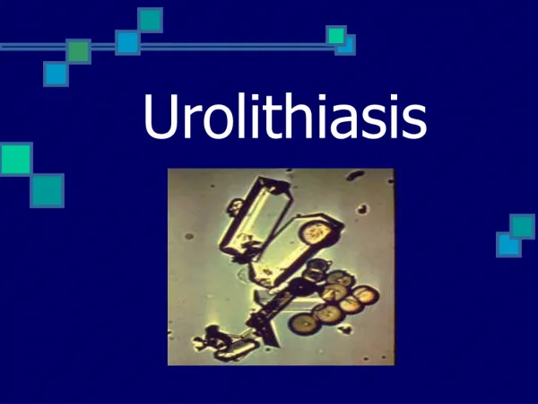
Urolithiasis. Key Points. Urolithiasis is the presence of calculi (stones) in the urinary tract. 75% are composed of calcium oxalate or calcium phosphate but may contain other substances such as uric acid.

E N D
Key Points • Urolithiasisis the presence of calculi (stones) in the urinary tract. • 75% are composed of calcium oxalate or calcium phosphate but may contain other substances such as uric acid. • A diet high in calcium is not believed to increase the risk of stone formation unless there is a preexisting metabolic disorder or renal tubular defect. • Reoccurrence is increased (35 to 50%) with family history, or first onset before age of 25 years. • Most clients can expel stones without invasive procedures. Factors that influence whether a stone will pass spontaneously or not include the composition, size, and location of the stone.
Key Factors • The cause of urolithiasis is unknown. • There is an increased incidence of urolithiasis in males. • Urolithiasis formation is associated with: • Slow urine flow with supersaturation of urine • Damage to the lining of the urinary tract. • Decreased inhibitor substances in the urine. • Metabolic defects including: Increased intestinal absorption or decreased renal excretion of calcium. • Increased oxalate production (genetic) or ingestion from foods. • Increased production or decreased clearance of purines (contributing to increased uric-acid levels). • High urine acidity or alkalinity contributes to stone formation. • Urinary stasis, urinary retention, immobilization, and dehydration contribute to an environment favorable for stone formation.
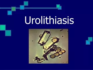
Diagnostic Procedures and Nursing Interventions Expected Findings • Urinalysis; • Increased RBCs, WBCs, and bacteria from urinary stasis. • Increased urine turbidity and odor (if urine is infected). • Crystals noted on microscopic exam. • Abnormal serum calcium, phosphate, and uric-acid levels in the presence of metabolic disorders/defects. • Elevated WBC if infection is present. • Radiology examination • KUB (x-ray of kidney, ureters, bladder), • CT scan, or IVP (intravenous pyelogram) • CT or MRI is used to identify cystine or uric-acid stones, which cannot be seen on standard x-rays. A renal ultrasound or cystoscopy may confirm diagnosis. • US
Therapeutic Procedures and Nursing Interventions Non-surgical Management • Extracorporeal shock wave lithotripsy (ESWL) (conscious sedation and ECG monitoring during the procedure). • Surgical management • Stenting is the placement of a small tube in the ureter during ureteroscopy. • Retrograde ureteroscopy uses a basket, forceps, or loop on the end of the ureteroscope to grasp and remove the stone. • Percutaneous ureterolithotomy/nephrolithotomy. • Open surgery for large or impacted stones • Ureterolithotomy(into the ureter) • Pyelolithotomy(into the kidney pelvis) • Nephrolithotomy(into the kidney)
Nursing Interventions • Prepare the client for procedures. • Teach the client about the procedures. • Report abnormal findings to the provider. • Teach the client about ESWL. Strain urine following the procedure. • Bruising is normal at the site where waves are applied. • Provide preoperative and postoperative care as indicated.
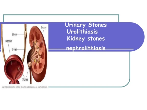
Assessments • Severe pain (renal colic) • Increases with stone movement. • Flank pain suggests stones in the kidney or ureter( if radiates to the abdomen, scrotum, testes, or vulva is suggestive of stones in the ureters or bladder). Nausea/vomiting • Urinary frequency or dysuria (occurs with stones in the bladder) • Pallor • Diaphoresis • Vital signs: Tachycardia, tachypnea, increased or decreased blood pressure with pain • Oliguria/anuria (occurs with stones that obstruct urinary flow) • Hematuria
NANDA Nursing Diagnoses • Acute pain • Risk for infection • Deficient knowledge • Risk for injury (renal) • Fear • Anxiety
Nursing Interventions • Assess/Monitor • Pain status • Intake and output • Urinary pH • Administer prescribed medications, such as: • Analgesics (opioids , NSAIDS ). • Spasmolytic drugs. • Antibiotics if infection is present. • Strain all urine to check for passage of the stone; save the stone for laboratory analysis.
Nursing Interventions • Encourage increased oral intake to 3,000 mL/day unless contraindicated. • Administer IV fluids as prescribed. • Encourage ambulation????. • Provide client education regarding the role of diet and medications in the treatment and prevention of urinary stones. • Calcium phosphate Dietary interventions Limit intake of food high in animal protein (Reduction of protein intake decreases calcium precipitation). • Limit sodium intake. • Reduced calcium intake (dairy products) is individualized. • Medications Thiazide diuretics (hydrochlorothiazide) to increase calcium reabsorption Orthophosphates to decrease urine saturation of calcium oxalate Sodium cellulose phosphate to reduce intestinal absorption of calcium
Nursing Interventions • Uric acid (urate) Dietary interventions Decrease intake of purine sources (organ meats, poultry, fish, gravies, sardines). • Medications to prevent formation of uric acid • Potassium or sodium citrate or sodium bicarbonate to alkalinize the urine
Complications and Nursing Implications • Treat an obstruction of the ureter and interruption of urinary flow as an emergency, and report it immediately. • Monitor for signs of infection, and report abnormal findings. • Monitor for signs of urine outflow obstruction (hydronephrosis) of the affected kidney, and report abnormal findings. • Renal failure (For information, refer to chapter 50, Renal Failure.)