Download
1 / 80
850 likes | 1.51k Views
Urolithiasis. Syndrome of swollen scrotum. Pavlo Hoschynsky. Urolithiasis. Introduction

E N D
Urolithiasis. Syndrome of swollen scrotum. Pavlo Hoschynsky
Urolithiasis Introduction Urolithiasis is increasingly recognized in pediatric patients and is encountered in a variety of clinical settings.The wide geographic variation in the incidence of urolithiasis in childhood is related to climatic, dietary, and socioeconomic factors. Approximately 7%of urinary calculi occur in children younger than 16 years of age.Many children with stone disease have a metabolic abnormality. Revolutionary advances in the minimally invasive and noninvasive management of stone disease over the past 2 decades have greatly facilitated the ease with which stones are removed. Given the frequency with which stones recur, the development of a medical prophylactic program to prevent stone recurrences is desirable.The lifetime prevalence of kidney stone disease is estimated at 1% to 15%, with the probability of having a stone varying according to age, gender, race, and geographic location. Stone disease typically affects boys more commonly as much as two to three times more frequently than females. Upper urinary tract stones occur more commonly in boys than girls by a ratio of 1.4:1 to 2.1:1.
Classification of stones Stone size: • <5 mm, • 5-10 mm, • > 10-20 mm, • > 20 mm.
Classification of stones Stone location: • upper calyx, • middle calyx or lower calyx, • renal pelvis, • upper ureter, • middle ureter or distal ureter, • urinary bladder.
Classification of stones X-ray characteristics
Calcium oxalate monohydrates • Calcium oxalate dihydrates
Diagnostic steps in urolithiasis (UTI urinary tract infection, CT computed tomography, MR1 magnetic resonance imaging, PTH parathyroid hormone, pC02 partial pressure of carbon dioxide)
Fig.a,b. A 17-year-old girl with cystinuria. a) Abdominal plain radiograph showing urolithiasis on the left, b) IVU showing hydronephrosis on the left due to urolithiasis
Fig. a,b. A 4-year-old boy with incomplete RTA and hyperoxaluria, a Sonogram of right kidney showing medullary nephrocalcinosis grade III (Dick et al. 1999). b Sonogram of bladder showing an ureteral stone on the right immediately before the ureterovesical junction
An 8-year-old boy with primary hyperparathyroidism, hypercalciuria, and urinary tract infection. Abdominal plain radiograph showing a huge ureteral stone on the left immediately before the ureterovesical junction
Bilateral Ureteric Calculus in a patient presenting with Anuria Helical or Spiral CT provides 3D reconstruction. Helical refers to path the X ray follows on Gantry. These are rapidly performed and do not require contrast agents for reconstruction.
Evaluation for a suspected stone. (RBUS)-renal/bladder ultrasound
extracorporeal shockwave lithotripsy (ESWL) percutaneous nephrolithotomy -(PCNL)
Recommendations for pain relief during renal colic: -1st choice: treatment should be started with an NSAID(Diclophenac sodium, Indomethacin, Ibuprofen) -2nd choice: Hydromorphine(Pentazocine,Tramadol) -Diclofenac sodium is recommended to counteract recurrent pain after an episode of ureteral colic For septic patients with obstructing stones, the collecting system should be urgently decompressed, using either percutaneous drainage or ureteral stenting. Definitive treatment of the stone should be delayed until sepsis is resolved. Medical expulsive therapy Alpha-blockers (Tamsulosin, 0.4 mg, doxazosin,terazosin, alfuzosin and naftopidil) Calcium-channel blockers(nifedipine) Corticosteroids Chemolytic dissolution of stones: -Percutaneous irrigation chemolysis -Oral Chemolysis
The figure shows a 12 month-old child treated with the Modulith SLK (Storz Medical AG, Kreuzlingen).
Operation: percutaneous nephrolithotomy ■ Rarely used in pediatric surgery ■ Utilize a nephroscope or ureteroscope ■ Extract with visualization ■ Break larger stones using ultrasonography Operation: open stone removal ■ Rarely necessary, only when urinary calculi are not amenable to ESWL or PL ■ Make an incision below the 12th rib ■ Expose the kidney and the ureter ■ Open the renal pelvis and extract the stone (or ureter in the case of a ureteral stone) ■ Wash the entire calyx system ■ Suture the pyelon or the ureter Postoperative care ■ Ureter drain for 2–5 days with an antegrade contrast X-ray before drain removal ■ Antibiotic therapy as prophylaxis in cases of vesicoureteral reflux ■ Urine culture once a month ■ Ultrasonography Prognosis ■ Stone recurrence is rare if urine is sterile and an obstruction does not occur
Scrotal Pain and Swelling Outline • Embryology and anatomy • Causes of Pain and Swelling • Torsion, Epididymitis, Orchitis, Trauma • History, Physical, Radiologic Exams, Labs • Causes of Swelling • Hydrocele, Varicocele, Spermatocele, Tumor, Idiopathic
Embryology • Descent of testes at 32-40 wks gestation • Descends within processes vaginalis • Outpouching of peritoneal cavity • Tunica vaginalis is potential space that remains after closure of process vaginalis
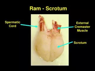
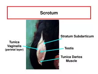
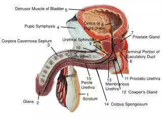
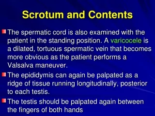
Anatomy • Spermatic cord –testicular vessels, lymph, vas deferens • Epididymis - sperm formed in testicle and undergo maturation, stored in lower portion • Vas Deferens – muscular action propels sperm up and out during ejaculation • Gubernaculum – fixation point for testicle to tunica vaginalis • Tunica Vaginalis – potential space • Encompasses anterior 2/3’s of testicle • Tunica albuginea is inner layer opposing testis
Anatomy – Nuts and Bolts Posterior Anterior
Causes of Pain and Swelling • Pain • Testicular torsion • Torsion of appendix testis • Epididymitis • Trauma • Orchitis and Others • Swelling • Hydrocele • Varicocele • Spermatocele • Tumor
Torsion • Inadequate fixation of testes to tunica vagnialis at gubernaculum • Torsion around spermatic cord • Venous compression to edema to ischemia
Epidemiology • Accounts for 30% of all acute scrotal swelling • Bimodal ages – neonatal (in utero) and pubertal ages • 65% occur in ages 12-18yo • Incidence 1 in 4000 in males <25yo • Increased incidence in puberty due to inc weight of testes
Predisposing Anatomy • Bell-clapper deformity • Testicle lacks normal attachment at vaginalis • Increased mobility • Tranverse lie of testes • Typically bilateral • Prevalence 1/125
Torsion: Clinical Presentation • Abrupt onset of pain – usually testicular, can be lower abdominal, inguinal • Often < 12 hrs duration • May follow exercise or minor trauma • May awaken from sleep • Cremasteric contraction with nocturnal stimulation in REM • Up to 8% report testicular pain in past
Torsion: Examination • Edematous, tender, swollen • Elevated from shortened spermatic cord • Horizontal lie common (PPV 80%) • Reactive hydrocele may be present • Cremasteric reflex absent in nearly all (unreliable in <30mo old) (PPV 95%) • Prehn’s sign elevation relieves pain in epididymitis and not torsion is unreliable
Intermittent Torsion • Intermittent pain/swelling with rapid resolution (seconds to minutes) • Long intervals between symptoms • PE: testes with horizontal lie, mobile testes, bulkiness of spermatic cord (resolving edema) • Often evaluation is normal – if suspicious need GU followup
Diagnosis – “Time is Testicle” • Ideally -- prompt clinical diagnosis • Imaging • Color doppler – decreased intratesticular flow • False + in large hydrocele, hematoma • Sens 69-100% and Spec 77-100% • Lower sensitivity in low flow pre-pubertal testes • Nuclear Technetium-99 radioisotope scan • Show testicular perfusion • 30 min procedure time • Sens and spec 97-100%
Acute torsion L testis • Dec blood flow on L • Late torsion on R • Inc blood flow around but dec flow w/in testis
Images - Torsion • Decreased echogenicity and size of right testicle • Nuclear medicine scan shows "rim sign“ =no flow to testicle and swelling
Management • Detorsion within 6hr = 100% viability • Within 12-24 hrs = 20% viability • After 24 hrs = 0% viability • Surgical detorsion and orchiopexy if viable • Contralateral exploration and fixation if bell-clapper deformity • Orchiectomy if non-viable testicle • Never delay surgery on assumption of nonviability as prolonged symptoms can represent periods of intermittent torsion
Manual Detorsion • If presents before swelling • Appropriate sedation • In 2/3rds of cases testes torses medially, 1/3rd lateral • Success if pain relief, testes lowers in scrotum • Still need surgical fixation
Torsion: Special Considerations • Adolescents may be embarrassed and not seek care until late in course • Torsion 10x more likely in undescended testicle • Suspicious if empty scrotum, inguinal pain/swelling • Adult Emergency Physicians accurate in bedside US diagnoses with sens of 95% and specificity of 94% (missed 1 epididymitis, no torsion) Blavis M., Emergency Evaluation of Patients Presenting with A Cute Scrotum, Academy of Emergency Medicine. Jan 2001
Neonatal Torsion • 70% prenatal, 30% post-natal • Post-natal typically 7-10 days after birth • Unrelated to gestation age, birth weight • Post-natal presents in typical fashion • Doppler U/S and radionucleotide scans less accurate with low blood flow in neonates • Surgical intervention if post-natal • Prenatal torsion presents with painless testicular swelling, rare testicular viability • Rare intervention in prenatal torsion
Torsion of Appendix Testis • Appendix testis • Small vestigial structure, remnant of Mullerium duct • Pedunculated, 0.3cm long • Other appendix structures • Prepubertal estrogen may enlarge appendix and cause torsion