Download
1 / 1
10 likes | 184 Views
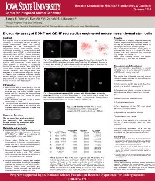
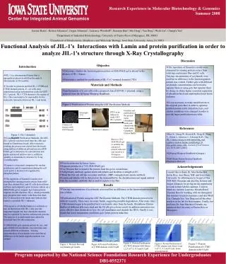
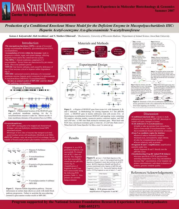
1. 2. 3.10.1.1. 2.3.1.78. 3.1.6.18. 3.2.1.31. 3.1.6.14. 7000. O. O. 1600. 3.2.1.50. 500. Materials and Methods. Step 1. Isolate developing embryo at blastocyst stage. This embryo is from a strain of mice with gray fur. .

E N D
1 2 3.10.1.1 2.3.1.78 3.1.6.18 3.2.1.31 3.1.6.14 7000 O O 1600 3.2.1.50 500 Materials and Methods Step 1. Isolate developing embryo at blastocyst stage. This embryo is from a strain of mice with gray fur. Step 2. Remove embryonic stem cells from gray-fur blastocyst. Grow stem cells in tissue culture. Embryonic stem cell generation and transfection (fig.4) Screening for knockout mice (fig.4) General Design and Strategy (Fig.3) PCR/Cloning (Fig.3a,3b) Exon 4 Exon 5 Exon 6 Exon 4 Exon 5 Exon 6 a) c) X X Step 3. Transfect stem cells with targeting vector construct. Select for homologous recombination by growing stem cells in neomycin and gancyclvir. PCR amplification of three fragments covering the region of interest Step 4. Remove homologously recombined stem cells from petri dish and inject into a new blastocyst that would have only white fur. d) TK LoxP Exon 5 Neo Homologous Recombination A B C FRT b) Exon 4 Exon 5 1348 bp Exon 6 3500 bp e) Neo Step 1 Step 1 Step 2 FRT LoxP Step 6. The progeny will be normal white fur mice but others will be chimeric mice. Chimeric mice have many of their cells from the original white fur blastocyst but some of their cells will be derived from recombinant stem cells. Step 2 Step 5. Implant several chimeric blastocysts into pseudo-pregnant, white fur mouse. Breeding with FLPe Mice PGK-Neo PGK-TK LoxP FRT LoxP Exon 4 Exon 5 Exon 6 f) pBY49a pBYLoxPa FRT LoxP 7474 bp 3004 bp Amp Amp Breeding with Tissue- Specific Cre Mice Step 7. Mate the chimeric mice with wild-type white fur mice. If the gonads of the chimeric mice were derived from recombinant stem cells, all the offspring will have gray fur. Every cell in gray mice are heterozygous for the homologous recombination. Step 8. Mate heterozygous gray mice (+/ H) and genotpye the gray offspring. Identify homozygous recombinants (H / H) and breed them to produce a strain of mice with both alleles knocked out. The pure breeding mouse strain is a "knockout mouse". Human Chromosome 8 Exon 4 Exon 6 g) LoxP Figure 4. Schematic of ES cell recombination and knockout mouse generation (work to be conducted by the University of Iowa Gene targeting core facility). Illustration from http://www.bio.davidson.edu/Courses/genomics/method/homolrecomb.html) Figure 1. Chromosomal location of the HGSNAT gene and predicted membrane topology for the N-acetyltransferase enzyme it codes for. Shown are the 11 trans-membrane domains of the protein (Fan et.al 2006). Discussion • Conditional knockout mice: a means to study pathogenesis by restricting MPS IIIC phenotype to certain cell types, lineages or tissues. • Cells deficient in N-acetyltransferase: • Unable to take up enzyme from extracellular fluid or from nearby N-acetyltransferase + cells, due to membrane-bound characteristic of enzyme. • Exon 5 as candidate region for deletion: • Removal induces frameshift mutation. • Isolated exon, surrounded by ample introns. • Codes for a transmembrane domain. • Occurs relatively early in protein. • Fragment B and C Amplification: amplification currently under way. • Insert fragments B and C directly into the pBY49a vector. • Troubleshooting-Frag BC initial PCR failures: • Increase specificity of PCR reaction (raise annealing temperature, lower Taq concentration, decrease Mg2+ concentration, lower template and primer concentrations), individual evaluation of primers, genomic template quality. Results a) b) 2 3 4 5 6 7 8 9 10 11 • Fragment A, as a PCR generated amplicon, was successfully TA cloned into pCR2.1 (data not shown). This fragment was then successfully cloned into the pBY49a vector (fig.5). • Amplification of fragment B and C has been unsuccessful using the following PCR primers (Table 1). New primers are currently being evaluated. H2CO S COOH H2COH Heparan N-Sulfatase O 1600 bp O O MPS IIIA O~ O NAc N S O S N-acetyltransferase Figure 5.a) Lane 1; NotI-HpaI digestion of the pBY49a vector (1).Lane 2: Gel isolated NotI-EcoCRI digestion of fragment A insert derived from its pCR2.1 parent. b)DNA digestions (BamHI and HincII) of colonies transformed with a ligation of pBY49a and Fragment A (lanes 2-11). Successful ligations should yield a 1219 bp fragment as seen in lanes 2, 7, and 9. MPS IIIC H2CO S COOH H2COH O O O O~ O N-acetyl-a-D-glucosaminidase NAc NAc O S MPS IIIB H2CO S COOH Name Primer Sequence Annealing Temperature O O O O~ NAc Fragment A- Forward 5’-ACAACTCTACT-GCAGAATGTCTGTCCCC-3’ 68˚C H2CO S Fragment A- Reverse 5’-TTTATTGTGTTGGGAA-GAGTCAGAGTCCACGTG-3’ 68˚C N-acetylglucosamine 6-sulfatase O O~ MPS IIID Fragment BC- Forward 5’-TTGTGGAGAGAAAGGA-AACTTTGAGGTTTGGGTT-3’ 64.4˚C NAc Fragment BC- Reverse 5’-GAAACTCACTCTGTAGA-CCAGGCTGGCCTCAAAC-3’ 64.4˚C Figure 2. Heparan Sulfate degradation pathway. Enzyme deficiencies at certain steps (shown as circle with slash) block pathway progression. Failure to acetylate the non-reducting terminal α-glucosamine residue results in MPS IIIC. Table 1. PCR primers used for amplification of fragments A and BC. • Research Experience in Molecular Biotechnology & Genomics • Summer 2007 Center for Integrated Animal Genomics Production of a Conditional Knockout Mouse Model for the Deficient Enzyme in Mucopolyaccharidosis IIIC: Heparin Acetyl-coenzyme A:α-glucosaminide N-acetyltransferase Kaizan J. Kalyaniwalla1, Rafi Awedikian2, and N. Matthew Ellinwood2. 1Biochemistry, University of Wisconsin-Madison, 2Department of Animal Science, Iowa State University • Introduction • The mucopolysaccharidoses (MPS): a group of lysosomal storage enzymopathies defined by glycosaminoglycan (GAG) accumumulation. • Accumulation of GAGs within the lysosome: leads to progressive cellular, organ, and system damage, affecting primarily bones and/or the central nervous system. • The MPSs: 7 clinical syndromes comprised of 11 enzymopathies. Severe forms are characterized by pre-mature death, often in the pediatric period. • MPS III: four known subtypes, each caused by deficiency of one of four enzymes critical to heparan sulfate degradation (fig.2). • MPS IIIC: autosomal recessive deficiency of a lysosomal membrane enzyme (heparin acetyl-coenzyme A:α-glucosaminide N-acetyltransferase (HGSNAT, N-acetyltransferase)). • To date no animal models of MPS IIIC exist for clinical and pathology investigations. Experimental Design Figure 3.a) Region of HGSNAT gene from exons 4-6, with fragments A, B, and C indicated. b) Methodology for cloning fragments A-C into pBY49a vector. c)HGSNAT gene in murine embryonic stem cells (exons 4-6). d) Homologous recombination between HGSNAT and targeting vector containing TK negative selection marker, neomycin positive selection marker, and FRT and LoxP sites. e) FRT sites flank neomycin resistance gene. When bred with FLPe mice, neocmycin resistance gene is removed. f) LoxP sites flank exon 5. When bred with Tissue Specific Cre Mice, exon 5 is removed (g). • N-acetyltransferase: • Last MPS causing gene to be mapped and characterized. • Unique: only non-hydrolytic, membrane-bound MPS associated enzyme. • Prototype of new class of enzyme that transport activated acetyl residues across cell membranes (Hrebicek et.al 2006). • MPS IIIC patients: carry out acetyl-CoA/CoA exchange, but no transfer of bound acetyl group to glucosamine (Ausseil et.al 2005). References/Acknowledgements Ausseil, J., et al., An acetylated 120-kDa lysosomal transmembrane protein is absent from mucopolysaccharidosis IIIC fibroblasts: a candidate molecule for MPS IIIC. Mol Genet Metab, 2006. 87(1): p. 22-31. Fan, X., et al., Identification of the gene encoding the enzyme deficient in mucopolysaccharidosis IIIC (Sanfilippo disease type C). Am J Hum Genet, 2006. 79(4): p. 738-44. Hrebicek, M., et al., Mutations in TMEM76* cause mucopolysaccharidosis IIIC (Sanfilippo C syndrome). Am J Hum Genet, 2006. 79(5): p. 807-19. Neufeld, E.F., Muenzer, J., The Mucopolysaccharidoses, in The Metabolic and Molecular Bases of Inherited Disease, C.R. Scriver, Beaudet, A. L., Sly, W. S., Valle D., Editor. 2001, McGraw-Hill, Health Professions Division: New York. p. 3421-3452. No author. 2002. Homolgous Recombination Method (and Knockout Mouse). <http://www.bio.davidson.edu/Courses/genomics/method/homolrecomb.html> Department of Biology, Davidson College, Davidson, North Carolina. Seyrantepe, V., et al. Lysosomal N-acetyltransferase deficient in mucoplysaccharidosis type IIIc is encoded by the TMEM76 gene on human chromosome 8. in 9th international Symposium on Mucopolysaccharide and related Diseases. 2006. Venice, Italy. I would like to personally thank Liz Snella, Mary Jane Long, and Stephanie Patokca for all their help around the lab. Special thanks to Rafi Awedikian for training me in the techniques of cloning and Matthew Ellinwood for his endless support. This work was supported in part by grants from the National MPS Society Inc., A Life for Elisa-Sanfilippo Children’s Research Foundation, and the NSF-REU research grant. Program supported by the National Science Foundation Research Experience for Undergraduates DBI-0552371