Download
1 / 1
10 likes | 86 Views
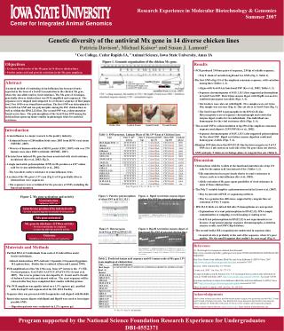
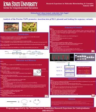
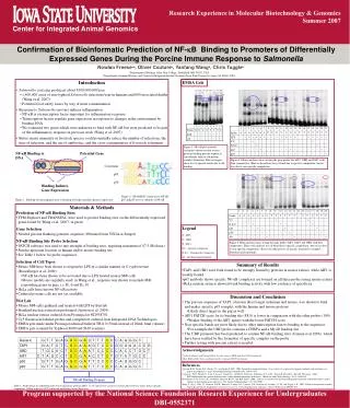
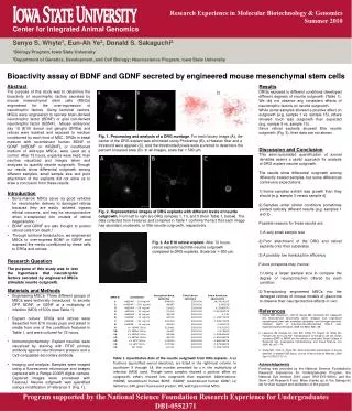
Research Experience in Molecular Biotechnology & Genomics Summer 2007. Center for Integrated Animal Genomics. Isolation and Purification of GST-GFP Protein for Screening Anti-GFP Antibody. Chioma Ebiringa 1 , Laurence Woodruff 2 , and Kristen Johansen 2

E N D
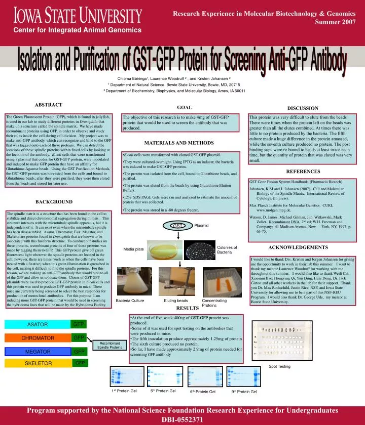
Research Experience in Molecular Biotechnology & Genomics • Summer 2007 Center for Integrated Animal Genomics Isolation and Purification of GST-GFP Protein for Screening Anti-GFP Antibody Chioma Ebiringa1, Laurence Woodruff 2 , and Kristen Johansen 2 1 Department of Natural Science, Bowie State University, Bowie, MD, 20715 2 Department of Biochemistry, Biophysics, and Molecular Biology, Ames, IA 50011 ABSTRACT GOAL DISCUSSION The Green Fluorescent Protein (GFP), which is found in jellyfish, is used in our lab to study different proteins in Drosophila that make up a structure called the spindle matrix. We have made recombinant proteins using GFP, in order to observe and study their roles inside the cell during cell division. My project was to make anti-GFP antibody, which can recognize and bind to the GFP that was tagged onto each of these proteins. We can detect the locations of these spindle proteins within fixed cells by looking at the location of the antibody. E.coli cells that were transformed using a plasmid that codes for GST-GFP protein, were inoculated and induced to make GFP protein that have an affinity for Glutathione Agarose beads. Using the GST Purification Methods, the GST-GFP protein was harvested from the cells and bound to Glutathione beads; after they were purified, they were then eluted from the beads and stored for later use. The objective of this research is to make 4mg of GST-GFP protein that would be used to screen the antibody that was produced. This protein was very difficult to elute from the beads. There were times when the protein left on the beads was greater than all the elutes combined. At times there was little to no protein produced by the bacteria. The fifth culture made a huge difference in the protein amassed, while the seventh culture produced no protein. The post binding sups were re-bound to beads at least twice each time, but the quantity of protein that was eluted was very small. MATERIALS AND METHODS • E.coli cells were transformed with cloned GST-GFP plasmid. • They were cultured overnight. Using IPTG as an inducer, the bacteria was induced to make GST-GFP proteins. • The protein was isolated from the cell, bound to Glutathione beads, and purified. • The protein was eluted from the beads by using Glutathione Elution Buffers. • 12% SDS PAGE Gels were ran and analyzed to estimate the amount of protein that was collected. • The protein was stored in a -80 degrees freezer. REFERENCES GST Gene Fusion System Handbook. (Pharmacia Biotech) Johansen, K.M and J. Johansen (2007). Cell and Molecular Biology of the Spindle Matrix. International Review of Cytology. (In press). Max Planck Institute for Molecular Genetics. CURL www.molgen.mpg.de. Watson, D. James, Michael Gilman, Jan Witkowski, Mark Zoller. Recombinant DNA, 2nd ed; W.H. Freeman and Company: 41 Madison Avenue, New York, NY, 1997; p. 63-75. BACKGROUND The spindle matrix is a structure that has been found in the cell to stabilize and direct chromosomal segregation during mitosis. This structure interacts with the microtubule spindle apparatus, but it is independent of it. It can exist even when the microtubule spindle has been disassembled. Asator, Chromator, East, Megator, and Skeletor are proteins found in Drosophila that are known to be associated with this fusiform structure. To conduct our studies on these proteins, recombinant proteins of four of these proteins was made by tagging them to GFP. This GFP protein give off green fluorescent light wherever the spindle proteins are located in the cell; however, there are times (such as when the cells have been treated with a fixative) when this green illumination is quenched in the cell, making it difficult to find the spindle proteins. For this reason, we are making an anti-GFP antibody that would bind to all of the GFP and allow us to locate them. Clones of GST-GFP plasmids were used to produce GST-GSP protein in E.coli cells and this protein was used to produce GFP antibody in mice. These mice are currently being screened to select the best responder for production of monoclonal antibodies. For this purpose, I am inducing more GST-GFP protein that would be used in screening the hybridoma lines that will be made by the Hybridoma Facility. GST Plasmid ACKNOWLEDGEMENTS Colonies of Bacteria Media plate I would like to thank Drs. Kristen and Jorgen Johansen for giving me the opportunity to work in their lab this summer. I want to thank my mentor Laurence Woodruff for working with me throughout this summer. I would also like to thank Weili Cai, Xiaomin Bao, Hongying Qi, Yun Ding, Huai Deng, Dr. Jack Girton and all other workers in the lab for their support. Thank you Dr. Max Rothschild, Justin Rice, NSF, and Iowa State University for allowing me to be a part of this NSF-REU Program. I would also thank Dr. George Ude, my mentor at Bowie State University. Bacteria Culture Eluting beads Concentrating Proteins RESULTS • At the end of five week 400ug of GST-GFP protein was produced. • Some of it was used for spot testing on the antibodies that were produced in mice. • The fifth inoculation produce approximately 1.25mg of protein • The sixth culture produced no protein. • So far, I have made approximately 2.9mg of protein needed for screening GFP antibody GFP ASATOR GFP CHROMATOR Recombinant Spindle Proteins MEGATOR GFP SKELETOR GFP Spot Testing 1st Protein Gel 5th Protein Gel 6th Protein Gel 9th Protein Gel Program supported by the National Science Foundation Research Experience for Undergraduates DBI-0552371