Download
1 / 1
10 likes | 97 Views
Research Experience in Molecular Biotechnology & Genomics Summer 2010. Center for Integrated Animal Genomics. Senyo S. Whyte 1 , Eun-Ah Ye 2 , Donald S. Sakaguchi 2 1 Biology Program, Iowa State University

E N D
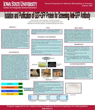
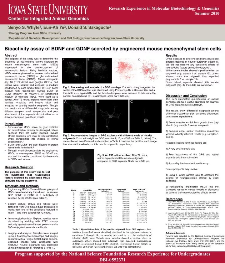
Research Experience in Molecular Biotechnology & Genomics • Summer 2010 Center for Integrated Animal Genomics Senyo S. Whyte1, Eun-Ah Ye2, Donald S. Sakaguchi2 1Biology Program, Iowa State University 2Department of Genetics, Development, and Cell Biology; Neuroscience Program, Iowa State University Bioactivity assay of BDNF and GDNF secreted by engineered mouse mesenchymal stem cells Abstract The purpose of this study was to determine the bioactivity of neurotrophic factors secreted by mouse mesenchymal stem cells (MSCs) engineered for the over-expression of neurotrophic factors. Using lentiviral vectors, MSCs were engineered to secrete brain-derived neurotrophic factor (BDNF) or glial cell-derived neurotrophic factor (GDNF). Mouse embryonic day 18 (E18) dorsal root ganglia (DRGs) and retinas were isolated and exposed to medium conditioned by each kind of MSC. DRGs in basal medium with recombinant human BDNF or GDNF (rhBDNF or rhGDNF), or conditioned medium of wild-type MSCs, were used as a control. After 72 hours, explants were fixed, their neurites visualized and images taken and analyzed to quantify neurite outgrowth. Though our results show differential outgrowth among different samples, small sample size and poor attachment of the explants did not allow us to draw a conclusion from these results. Results DRGs exposed to different conditions developed different degrees of neurite outgrowth (Table 1). We did not observe any consistent effects of neurotrophic factors on neurite outgrowth. While some samples showed a positive effect on outgrowth (e.g. sample 1 vs. sample 15), others showed much less outgrowth than expected (e.g. sample 5 vs. sample 15). Since retinal explants showed little neurite outgrowth (Fig. 3), their data are not shown. DRG Fig. 1. Processing and analysis of a DRG montage. For each binary image (A), the center of the DRG explant was eliminated using Photoshop (B), a Hessian filter and a threshold were applied (C), and the thresholded pixels were summed to determine the percent occupied area (D). In all images, scale bar = 500 μm. • Discussion and Conclusion • This semi-automated quantification of axonal densities seems a useful approach for analysis of DRG explant neurite outgrowth. • The results show differential outgrowth among differently treated samples, but some differences contravene expectations: • Some samples exhibit less growth than they should (e.g. sample 3 versus sample 4). • Samples under similar conditions sometimes yielded radically different results (e.g. samples 1 and 2). Possible reasons for these results are: • A very small sample size • Poor attachment of the DRG and retinal explants onto their substrates 3) A possibly low transduction efficiency • Future prospects may involve: • Using a larger sample size to compare the degree of neuroprotection offered by each condition • Transplanting engineered MSCs into the damaged retinas of mouse models of glaucoma to observe their neuroprotective effects in vivo • Introduction • Bone-marrow MSCs serve as good vehicles for neurotrophin delivery to damaged retinas because they are easily isolated, bypass ethical concerns, and may be neuroprotective when transplanted into models of retinal degeneration.1 • BDNF and GDNF are also thought to protect retinal cells from death.2 • Through lentiviral transduction, we engineered MSCs to over-express BDNF or GDNF and exposed the media conditioned by these cells to DRGs and retinas. Fig. 2. Representative images of DRG explants with different levels of neurite outgrowth. From left to right are DRG samples 1, 13, and 5 (from Table 1, below). The data collected from FeatureJ and compiled in Table 1 confirms the fact that each image has abundant, moderate, or little neurite outgrowth, respectively. Fig. 3. An E18 retinal explant. After 72 hours, retinal explants had little neurite outgrowth compared to DRG explants. Scale bar = 500 μm. Research Question The purpose of this study was to test the hypothesis that neurotrophic factors secreted by engineered MSCs stimulate neurite outgrowth. • Materials and Methods • Engineering MSCs: Three different groups of MSCs were lentivirally transduced to secrete GFP, BDNF, or GDNF at a multiplicity of infection (MOI) of 500x (see Table 1). • Explant culture: DRGs and retinas were dissected from E18 mouse pups and plated in media from one of the conditions featured in Table 1, and were cultured for 72 hours. • Immunocytochemistry: Explant neurites were visualized by staining with RT97 primary antibody against neurofilament proteins and a Cy3-conjugated secondary antibody. • Imaging and analysis: Samples were imaged using a fluorescence microscope and images captured with a Retiga 2000R digital camera. Captured images were processed with FeatureJ. Neurite outgrowth was quantified using a modification of reference 3 (Fig. 1). References Harper MM, Adamson L, Blits B, Bunge MB, Grozdanic SD, Sakaguchi DS. Brain-derived neurotrophic factor released from engineered mesenchymal stem cells attenuates glutamate- and hydrogen peroxide-mediated death of staurosporine-differentiated RGC-5 cells. Experimental Eye Research. 2009 Oct;89(4):538—48. Lawrence JM, Keegan DJ, Muir EM, Coffey PJ, Rogers JH, Wilby MJ, Fawcett JW, and Lund RD. Transplantation of Schwann cell line clones secreting GDNF or BDNF into the retinas of dystrophic Royal College of Surgeons rats. Investigative Ophthalmology and Visual Science, Jan 2004; 45: 267—74. Grider MH, Chen Q, Shine HD. Semi-automated quantification of axonal densities in labeled CNS tissue. Journal of Neuroscience Methods. 2006 Sep 15;155(2):172—9. Table 1. Quantitative data of the neurite outgrowth from DRG explants. Area fractions (quantified axonal densities), are listed in the rightmost column. In conditions 9 through 14, the number preceded by x is the multiplicity of infection (MOI) used. Though some samples showed a positive effect on outgrowth, others showed less outgrowth than expected. Abbreviations: rhBDNF, recombinant human BDNF; rhGDNF, recombinant human GDNF; LV, lentivirus; GFP, green fluorescent protein; Wt, wild type normal MSCs. Acknowledgements Funding was provided by the National Science Foundation’s Research Experience for Undergraduates Program, the National Eye Institute (NIH; grant 1R01EY019294), and the Stem Cell Research Fund. Many thanks go to the Sakaguchi lab for their support and facilitation of this project. Program supported by the National Science Foundation Research Experience for Undergraduates DBI-0552371