Download
1 / 10
100 likes | 194 Views
PNS and Transmission. February 09, 2010. PNS. Composed of neurons and ganglia. Ganglia are swellings associated with nerves that contain collections of cell bodies. Somatic division: serves the skin, skeleton, and tendons.

E N D
PNS and Transmission February 09, 2010
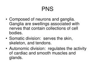
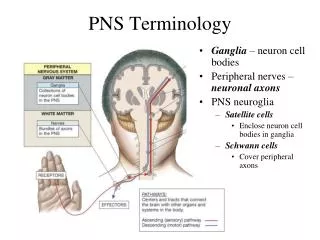
PNS • Composed of neurons and ganglia. Ganglia are swellings associated with nerves that contain collections of cell bodies. • Somatic division: serves the skin, skeleton, and tendons. • Autonomic division: regulates the activity of cardiac and smooth muscles and glands.
Types of PNS Nerves • Cranial: 12 pairs; many belong to the somatic division; includes the vagus nerve which has branches to most of the internal organs. • Spinal: 31 pairs; associated with the 3 regions of the vertebral column;
Somatic Division • Most actions are voluntary which means they originate in the cerebral cortex. • Others are reflexes: cranial (blinking) and spinal reflexes (hand on stove).
Autonomic Division • Sympathetic: most arise from the lower thoracic or lumbar region. Highly involved in the fight or flight reflex. • Parasympathetic: Craniosacral; promotes all the internal responses we associated with a relaxed state. • Commonalities: 1) they function automatically and usually involuntary, 2) they innervate all internal organs, and 3) they utilize 2 motor neurons and 1 ganglion for each impulse.
Nerve Impulses • Resting Potential: membrane is polarized (outside + inside -). The sodium potassium pumps are responsible for setting this up. • Action Potential: 1) depolarization (inside +); 2) repolarization (inside -). • If an axon is myelinated, the action potentials are stimulated between the nodes of Ranvier (faster potential) in non-myelinated it stimulates another part of the axomembrane. • All or None event. One way from cell body to axion terminal.
Transmission • Every axon terminates in an axon terminal. All of these lie close to a dendrite or the cell body of another neuron. • Pre-synaptic and Postsynaptic region. Between them is the Synaptic cleft.
Transmission • Transmission is carried out by molecules called neurotransmitters. These are stored in vesicles in the axon terminals. • Impulse reaches terminal opens calcium channels Calcium enters the terminal vesicles move toward membrane for exocytosis neurotransmitters are released and diffuse through synaptic cleft neurotransmitters bind with receptors on postsynaptic membrane. • Depending on the neurotransmitter and receptor the response will be excitation or inhibition.
Integration • Neurons can have many dendrites and can synapse with many other neurons. • An excitatory NT produces a potential change called a signal. The signal drives the polarity of a neuron closer to an action potential. An inhibitory NT does the opposite. • Integration is the summing up of all of the excitatory and inhibitory signals. Which ever side wins determines if an Action Potential will be transmitted.