Download
1 / 19
190 likes | 316 Views
Class 3 Ab + L AbL Review d/ dt [ AbL ] = k on [ Ab ][L] – k off [ AbL ] k off [1/s], k on [1/ Ms ], k off / k on = K D [M] why the lower the K D the tighter the binding? typical values for K D , k on , k off for Ab binding L

E N D
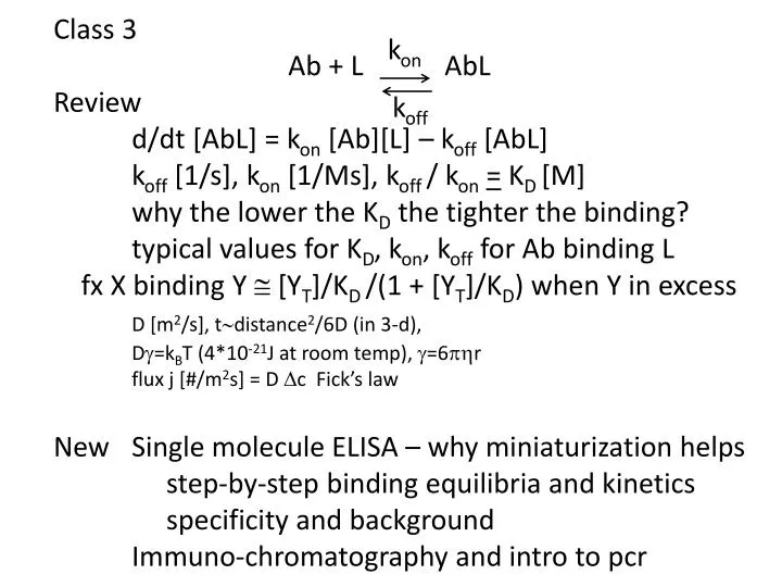
Class 3 Ab+ L AbL Review d/dt [AbL] = kon [Ab][L] – koff [AbL] koff [1/s], kon[1/Ms], koff/ kon= KD [M] why the lower the KD the tighter the binding? typical values for KD, kon, koff for Ab binding L fx X binding Y @ [YT]/KD /(1 + [YT]/KD) when Y in excess D [m2/s], t~distance2/6D (in 3-d), Dg=kBT (4*10-21J at room temp), g=6phr flux j [#/m2s] = D Dc Fick’s law New Single molecule ELISA – why miniaturization helps step-by-step binding equilibria and kinetics specificity and background Immuno-chromatography and intro to pcr kon koff
Why does miniaturization help? Enzyme catalytic rate kcat~100 substrate molecules/s per enzyme molecule Need threshold conc of dye ~100nM to see it the smaller the vol, the higher the conc how many molecules/50fl for 100nM? how many can one enz. molecule make in 30s? what are dimensions of 50fl volume fluorescence = advantage = 6*1023-7-15*50=3*103 [50x10-15*10-3m3 ]@ (4mm)3 emit longer wavelength than absorbed can block bright excitation light that would otherwise -> signal
Sensitivity of SiMoA Nat Biotech 28, 598 (2010) LOD std assay Why measure PSA? Why might it be useful to measure [PSA] after prost-x? Why might it not be useful?
Fig. 2A J Immunol Methods 378:102 (2012) given [AbT] @ 2nM [AbL]2 - [AbL] {[LT] + [AbT] + KD} + [LT] [AbT] = 0 or, since Ab in excess, [AbL]/[LT] @ [AbT]/KD / (1 + [AbT]/KD)
Fig.3 J Immunol Methods 378:102 (2012) The one in excess Is Ab or L in excess? Then t@ koff-1 / (1 + [AbT]/KD) Simplify [AbT]/KD Do results agree with figure? Are reactions B, C near equilibrium at 1000s? * What is equil [AbL]/[LT]? Does this agree with * ?
Fig. 4 J Immunol Methods 378:102 (2012) assumes KD of interaction = 1nM What step of rxn is this modeling? Why so flat? For which symbols is detection Ab in excess? Then fxAbL in complex @[DetAbT]/KD / (1 + [DetAbT]/KD) independent of [AbL]
Fig. 5 J Immunol Methods 378:102 (2012) What step is this modeling? What is the KD of this interaction? What is in excess? What is formula? Does this explain why almost all the points are 100%?
Specificity and background Background can come from non-tgt protein in sample or signal-generating species (e.g. enzyme) How many specific binding steps are there in label-free assay, like SPR? Suppose capture Ab binds tgt protein 107x more tightly (in terms of KD) than it binds high conc protein like albumin in biological sample [alb]plasma@mM What fx of capture Ab binds albumin? KD @ 10-9+7, so [alb]/KD / (1 + [alb]/KD) @ 0.1
What would this do in SPR assay? What would happen in sandwich assay? What would be [alb] after it is captured (wrongly) by capture Ab? If detectionAb also binds albwith mM affinity, what fraction of capture Ab-alb complexes bind detection Ab? If capture Ab is nM in device , [cap Ab-alb] ~0.1nM If [detAb] = nM, it is in excess over bound albumin, so f @ [detAb]/KD /(1 + [detAb]/KD) = 10-7 so 1011 capture Ab to start would -> 104detAb from alb
This is likely << threshold for signal in standard ELISA, but would -> signal in SiMoA Background in SiMoA was ~1% of beads Pcr is similar to sandwich immunoassays in that 2 specific binding events are required for signal (exponential amplification of tgt DNA) Biology often uses binding of multiple proteins to increase specificity of reactions
Lateral flow immunochromatography assays Common format for home tests (e.g. HCG - pregnancy) and now many medical lab tests DetAb on membrane but not attached; gets picked up and carried along by sample immobil. capt.Ab Neg control
Questions – how many gold particles at what density for easily visible line? why are they visible – ? just concentration or something special about plasmon resonance,which gives them color Flipped left-right from schematic; don’t know why C and T are diff. colors
Polymerase chain reaction = method to replicate piece of DNA “exponentially” 1. DNA polymeraseN strands are “anti-parallel” adds bases to 3’-end pol requires primer to start synthesis primer template
Short pieces of DNA (“oligo”nucleotides~1-100 bases) can be chemically synthesizedN, commercially available for <$1/base for 100nmol = 6x10¹⁶molecules, serve as “primers” to start DNA synthesis at particular place on DNA template
Pcr amplification of DNA using thermostable DNA polymerase (e.g. Taqpol) copy strand (B) forward primer Taqpol template strand (A) Melt DNA (94oC), cool (60oC) to anneal primers, extend (72oC) new strand A reverse primer
Old and new templates are not destroyed by melting, so repeated cycles of melting and polymerization -> 1 -> 2 -> 4 -> 8 -> … -> 2n copies of DNA region lying between 2 primer-annealing sites on initial template Polymerase copies ~1000b/min => ~1 hour for 230 =1010-fold amp. of a kb piece of DNA http://www.youtube.com/watch?v=_YgXcJ4n-kQ http://www.dnalc.org/ddnalc/resources/animations.html
Note target DNA seq., to ends of which oligos bind in right orientation, is amplified exponentially 2N, (N=#cycles) Non-tgt DNA, to which oligos may bind weakly, may be copied but only increases arithmetically ~N Different from signal amplification, e.g. use of enzymes that generate multiple fluors or dyes from each bound ligand in ELISA; here you make multiple copies of DNA ligand if there is 1 present to start with
Next week – first homework due Will begin paper that considers effects of flow and diffusion in sensors that use microfluidics – see Blackboard Considers “simple” revisions to model of binding kinetics when analyte concentrations can’t be considered spatially uniform