Download

1 / 29
290 likes | 650 Views
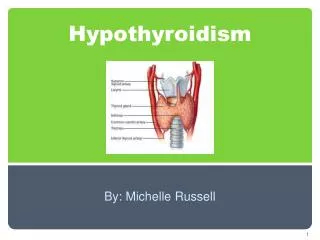
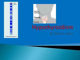
Hypothyroidism. Iodine deficiency remains the most common cause of hypothyroidism worldwide . In areas of iodine sufficiency, autoimmune disease (Hashimoto's thyroiditis) and iatrogenic causes (treatment of hyperthyroidism) are most common. Causes of Hypothyroidism. Primary

E N D
Hypothyroidism • Iodine deficiency remains the most common cause of hypothyroidism worldwide. • In areas of iodine sufficiency, autoimmune disease (Hashimoto's thyroiditis) and iatrogenic causes (treatment of hyperthyroidism) are most common .
Causes of Hypothyroidism Primary Autoimmune hypothyroidism: Hashimoto's thyroiditis, atrophic thyroiditis Iatrogenic: 131I treatment, subtotal or total thyroidectomy, external irradiation Drugs: iodine excess (including iodine-containing contrast media and amiodarone), lithium, antithyroid drugs, p-aminosalicylic acid, interferon- and sunitinib Congenital hypothyroidism: absent or ectopic thyroid gland, dyshormonogenesis, TSH-R mutation Iodine deficiency Infiltrative disorders: amyloidosis, sarcoidosis, hemochromatosis Overexpression of type 3 deoiodinase in infantile hemangioma Transient Silent thyroiditis, including postpartum thyroiditis Subacute thyroiditis Withdrawal of thyroxine treatment in individuals with an intact thyroid After 131I treatment or subtotal thyroidectomy for Graves' disease
Causes of Hypothyroidism Secondary • Hypopituitarism: tumors, pituitary surgery or irradiation, infiltrative disorders, Sheehan's syndrome, trauma, genetic forms of combined pituitary hormone deficiencies • Isolated TSH deficiency or inactivity • Bexarotene treatment • Hypothalamic disease: tumors, trauma, infiltrative disorders, idiopathic
Congenital Hypothyroidism Prevalence Hypothyroidism occurs in about 1 in 4000 newborns. It may be transient, especially if the mother has TSH-R blocking antibodies or has received antithyroid drugs, but permanent hypothyroidism occurs in the majority. Neonatal hypothyroidism is due to thyroid gland dysgenesis in 80–85%, to inborn errors of thyroid hormone synthesis in 10–15%, and is TSH-R antibody-mediated in 5% of affected newborns. The developmental abnormalities are twice as common in girls.
Congenital Hypothyroidism • <10% are diagnosed based on clinical features • prolonged jaundice, feeding problems, hypotonia, enlarged tongue, delayed bone maturation, and umbilical hernia. Importantly, permanent neurologic damage results if treatment is delayed. • neonatal screening programs have been established. These are generally based on measurement of TSH or T4 levels in heel-prick blood specimens. • When the diagnosis is confirmed, T4 is instituted at a dose of 10–15u g/kg per day, and the dose is adjusted by close monitoring of TSH levels.
Autoimmune Hypothyroidism Classification • Autoimmune hypothyroidism may be associated with a goiter (Hashimoto's thyroiditis) • atrophic thyroiditis Prevalence annual incidence rate of autoimmune hypothyroidism is up to 4 per 1000 women and 1 per 1000 men mean age at diagnosis is 60 years • subclinical hypothyroidism Subclinical hypothyroidism is found in 6–8% of women (10% over the age of 60) and 3% of men. The annual risk of developing clinical hypothyroidism is about 4% when subclinical hypothyroidism is associated with positive TPO antibodies. • symptoms become more readily apparent at this stage (usually TSH >10 mIU/L), which is referred to as clinical hypothyroidism or overt hypothyroidism.
Pathogenesis • In Hashimoto's thyroiditis, there is a marked lymphocytic infiltration of the thyroid with germinal center formation, atrophy of the thyroid follicles accompanied by oxyphilmetaplasia, absence of colloid, and mild to moderate fibrosis. • In atrophic thyroiditis, the fibrosis is much more extensive, lymphocyte infiltration is less pronounced, • susceptibility to autoimmune hypothyroidism is determined by a combination of genetic and environmental factors, and the risk of either autoimmune hypothyroidism or Graves' disease • HLA-DR polymorphisms are the best documented genetic risk factors for autoimmune hypothyroidism, especially HLA-DR3, -DR4, and -DR5 in Caucasians • CTLA-4, a T cell–regulatory gene, • Thyroid cell destruction is primarily mediated by the CD8+ cytotoxic T cells, which destroy their targets by either perforin-induced cell necrosis or granzyme B–induced apoptosis. In addition, local T cell production of cytokines, such as tumor necrosis factor (TNF), IL-1, and interferon (IFN-), • Up to 20% of patients with autoimmune hypothyroidism have antibodies against the TSH-R, which, in contrast to TSI, do not stimulate the receptor but prevent the binding of TSH. These TSH-R-blocking antibodies, therefore, cause hypothyroidism and, especially in Asian patients, thyroid atrophy • Rarely, patients have a mixture of TSI and TSH-R-blocking antibodies, and thyroid function can oscillate between hyperthyroidism and hypothyroidism as one or the other antibody becomes dominant.
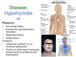
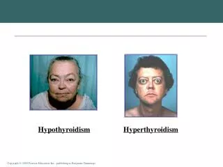
Clinical Manifestations • The onset is usually insidious • The skin is dry, and there is decreased sweating, thinning of the epidermis, and hyperkeratosis of the stratum corneum. Increased dermal glycosaminoglycan content traps water, giving rise to skin thickening without pitting (myxedema). • Typical features include a puffy face with edematous eyelids and nonpittingpretibial edema . There is pallor, often with a yellow tinge to the skin due to carotene accumulation. • Nail growth is retarded, and hair is dry, brittle, difficult to manage, and falls out easily. In addition to diffuse alopecia, there is thinning of the outer third of the eyebrows, although this is not a specific sign of hypothyroidism.
Signs and Symptoms of Hypothyroidism Symptoms • Tiredness, weakness • Dry skin • Feeling cold • Hair loss • Difficulty concentrating and poor memory • Constipation • Weight gain with poor appetite • Dyspnea • Hoarse voice • Menorrhagia (later oligomenorrhea or amenorrhea) • Paresthesia • Impaired hearing
Signs and Symptoms of Hypothyroidism Signs • Dry coarse skin; cool peripheral extremities • Puffy face, hands, and feet (myxedema) • Diffuse alopecia • Bradycardia • Peripheral edema • Delayed tendon reflex relaxation • Carpal tunnel syndrome • Serous cavity effusions
Treatment: Hypothyroidism • If there is no residual thyroid function, the daily replacement dose of levothyroxine is usually 1.6 ug/kg body weight (typically 100–150ug) • In the elderly, especially patients with known coronary artery disease, the starting dose of levothyroxine is 12.5–25 ug/d with similar increments every 2–3 months until TSH is normalized. • goal of treatment being a normal TSH, ideally in the lower half of the reference range • TSH responses are gradual and should be measured about two months after instituting treatment or after any subsequent change in levothyroxine dosage. • The clinical effects of levothyroxine replacement are slow to appear. Patients may not experience full relief from symptoms until 3–6 months after normal TSH levels are restored. • Adjustment of levothyroxine dosage is made in 12.5- or 25-ug increments if the TSH is high; decrements of the same magnitude should be made if the TSH is suppressed. • The use of levothyroxine combined with liothyronine (t3) ,, has not been confirmed in prospective studies
In patients of normal body weight who are taking 200ug of levothyroxine per day, an elevated TSH level is often a sign of poor adherence to treatment. • Such patients often have normal or high unbound T4 levels, despite an elevated TSH, because they remember to take medication for a few days before testing; this is sufficient to normalize T4, but not TSH levels. • Because T4 has a long half-life (7 days), patients who miss a dose can be advised to take two doses of the skipped tablets at once. • Other causes of increased levothyroxine requirements must be excluded, particularly malabsorption (e.g., celiac disease, small-bowel surgery), estrogen therapy, and drugs that interfere with T4 absorption or clearance such as cholestyramine, ferrous sulfate, calcium supplements, lovastatin, aluminum hydroxide, rifampicin, amiodarone, carbamazepine, and phenytoin.
Subclinical Hypothyroidism subclinical hypothyroidism refers to biochemical evidence of thyroid hormone deficiency in patients who have few or no apparent clinical features of hypothyroidism. There are no universally accepted recommendations for the management of subclinical hypothyroidism, but the most recently published guidelines do not recommend routine treatment when TSH levels are below 10 mU/L. It is important to confirm that any elevation of TSH is sustained over a 3-month period before treatment is given. As long as excessive treatment is avoided, there is no risk in correcting a slightly increased TSH. Moreover, there is a risk that patients will progress to overt hypothyroidism, particularly when the TSH level is elevated and TPO antibodies are present. Treatment is administered by starting with a low dose of levothyroxine (25–50 ug/d) with the goal of normalizing TSH. If thyroxine is not given, thyroid function should be evaluated annually.
Myxedema coma Myxedema coma still has a high mortality rate, Clinical manifestations include • reduced level of consciousness, seizures, • Hypothermia can reach 23°C (74°F). There may be a history of treated hypothyroidism with poor compliance, or the patient may be previously undiagnosed. Myxedema coma almost always occurs in the elderly and is usually precipitated by factors that impair respiration, such as drugs (especially sedatives, anesthetics, antidepressants), pneumonia, congestive heart failure, myocardial infarction, gastrointestinal bleeding, or cerebrovascular accidents. Sepsis should also be suspected. Exposure to cold may also be a risk factor. Hypoventilation, leading to hypoxia and hypercapnia, plays a major role in pathogenesis; hypoglycemia and dilutionalhyponatremia also contribute to the development of myxedema coma.
Myxedema coma • Levothyroxine can initially be administered as a single IV bolus of 500 ug, which serves as a loading dose. ,continued at a dose of 50–100 ug/d. • If suitable IV preparation is not available, the same initial dose of levothyroxine can be given by nasogastric tube (though absorption may be impaired in myxedema). • An alternative is to give liothyronine (T3) intravenously or via nasogastric tube, in doses ranging from 10 to 25 ug every 8–12 h. This treatment has been advocated because T4 T3 conversion is impaired in myxedema coma. However, excess liothyronine has the potential to provoke arrhythmias. • Another option is to combine levothyroxine (200 ug) and liothyronine (25u g) as a single, initial IV bolus followed by daily treatment with levothyroxine (50–100 ug/d) and liothyronine (10 ug every 8 h). • External warming is indicated only if the temperature is <30°C, as it can result in cardiovascular collapse . Space blankets should be used to prevent further heat loss. • Parenteral hydrocortisone (50 mg every 6 h) should be administered, because there is impaired adrenal reserve in profound hypothyroidism. • Any precipitating factors should be treated, including the early use of broad-spectrum antibiotics, pending the exclusion of infection. • Ventilatory support with regular blood gas analysis is usually needed during the first 48 hours. • Hypertonic saline or IV glucose may be needed if there is severe hyponatremia or hypoglycemia; hypotonic IV fluids should be avoided because
Iatrogenic hypothyroidismis a common cause of hypothyroidism and can often be detected by screening before symptoms develop. In the first 3–4 months after radioiodine treatment, transient hypothyroidism may occur due to reversible radiation damage. Low-dose thyroxine treatment can be withdrawn if recovery occurs. Because TSH levels are suppressed by hyperthyroidism, unbound T4 levels are a better measure of thyroid function than TSH in the months following radioiodine treatment. Mild hypothyroidism after subtotal thyroidectomy may • Secondary hypothyroidismis usually diagnosed in the context of other anterior pituitary hormone deficiencies; isolated TSH deficiency is very rare . TSH levels may be low, normal, or even slightly increased in secondary hypothyroidism; the latter is due to secretion of immunoactive but bioinactive forms of TSH. The diagnosis is confirmed by detecting a low unbound T4 level. The goal of treatment is to maintain T4 levels in the upper half of the reference range, because TSH levels cannot be used to monitor therapy.