Download
1 / 43
430 likes | 443 Views
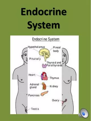
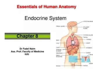
Chapter 36: Endocrine Control. Fig. 36-1b, p.618. Endocrine System. Main Sources Pituitary gland Adrenal glands Thyroid gland Parathyroid glands Pineal gland Thymus gland. Figure 36.2 Page 621. hypothalamus (part of the brain). pineal gland. pituitary gland, anterior lobe

E N D
Endocrine System Main Sources • Pituitary gland • Adrenal glands • Thyroid gland • Parathyroid glands • Pineal gland • Thymus gland Figure 36.2 Page 621
hypothalamus (part of the brain) pineal gland pituitary gland, anterior lobe pituitary gland, posterior lobe thyroid gland parathyroid glands (four) thymus gland adrenal gland (one pair) medulla pancreatic islets ovaries (one pair of female gonads) testes (one pair of male gonads) Fig. 36-2a, p.621
Hormones • Secreted by endocrine glands, endocrine cells, and certain neurons • Travel through the bloodstream to nonadjacent target cells
Hormone Action • Activation of receptor • Transduction of signal • Functional response
Responses to Hormones Vary • Different hormones activate different responses in the same target cell • Not all types of cells respond to a particular hormone
Two Main Hormone Types • Steroid hormones • Derived from cholesterol • Estrogens, progestins, androgens, cortisol, aldosterone • Peptide hormones • Peptides, proteins, or glycoproteins • Glucagon, ADH, oxytocin, TRH, insulin, somatotropin, prolactin, FSH, LH, TSH
Steroid Hormones hormone • Most diffuse across the plasma membrane and bind to a receptor • Hormone-receptor complex acts in nucleus to inhibit or enhance transcription receptor hormone-receptor complex Figure 36.3 Page 623 gene product
Protein Hormone glucagon • Hormone binds to a receptor at cell surface • Binding triggers a change in activity of enzymes inside the cell glucagon receptor cyclic AMP + Pi ATP cAMP activates protein kinase A Protein kinase A converts phosphorylase kinase to active form and inhibits an enzyme required for glucagon synthesis. Figure 36.3b Page 623
The Hypothalamus • Region in the forebrain • Contains hormone-secreting cells • Interacts with pituitary hypothalamus pituitary gland
Pituitary Gland • Pea-sized gland at base of hypothalamus • Two lobes • Posterior lobe stores and secretes hormones synthesized in the hypothalamus • Anterior lobe produces and secretes its own hormones
Posterior Lobe cell body in hypothalamus • Antidiuretic hormone (ADH) • Oxytocin (OCT) axons to the general circulation Figure 36.4 Page 624
Anterior Pituitary • ACTH • TSH • FSH • LH • PRL • STH Figure 36.5 Page 625
Normal Hormone Production • Generally, the body produces only very small amounts of hormones • To isolate 1 milligram of TRH, researchers dissected 7 metric tons of hypothalamic tissue
Abnormal Somatotropin Output • Gigantism • Pituitary dwarfism • Acromegaly
Thyroid Gland epiglottis thyroid cartilage (Adam’s apple) pharynx thyroid gland trachea (windpipe) parathyroid gland anterior posterior Fig. 36-6, p.626
Feedback Mechanisms • Negative feedback • Increase in hormone triggers activities that inhibit further secretion • Positive feedback • Increase in hormone triggers activities that stimulate further secretion
Stimulus Response + Blood level of thyroid hormone falls below a set point. – Hypothalamus TRH – Anterior Pituitary Rise in the blood level of thyroid hormone inhibits secretion of TRH and TSH. TSH Thyroid Gland Thyroid hormone is secreted Fig. 36-7, p.626
EXAMPLE: Cortisol • Cortisol secretion • Inhibits blood glucose uptake by muscle and other tissues • Causes breakdown of proteins to amino acids and conversion to glucose • Causes degradation of adipose tissue to fatty acids for use as energy source
Feedback Control of Cortisol Secretion • Hypothalamus senses rise in glucose and secretes less releasing hormone (CRH) • Anterior pituitary responds by secreting less ACTH • Adrenal cortex slows its secretion of cortisol
Localized Feedback in Adrenal Medulla • Norepinephrine secreted by neurons accumulates in the synaptic gap • Some molecules bind to receptors on the axon endings that secreted them • Prevents further secretion of norepinephrine by that axon
Thyroid Gland Disorders • Goiter • Hyperthyroidism • Hypothyroidism Figure 36.6 Page 626
EXAMPLE: Calcium Regulation • Parathyroid hormone (PTH) is the main regulator of calcium in the blood • It is secreted when calcium levels drop • PTH causes bone cells to digest bone tissue and release calcium • PTH also stimulates calcium reabsorption by the kidneys and absorption by the gut
EXAMPLE: Deformed Frogs • Something in water triggers deformities • Problem thyroid function? • Tadpoles from “hotspots” developed normally when given extra thyroid hormones • UV, parasites also play a role
Control of Glucose Metabolism insulin Glucose uptake Glucose to glycogen Glucose falls Glucose is absorbed Cells use glucose Glucose rises Glycogen to glucose glucagon
Diabetes Mellitus Excess glucose accumulates • Type 1 • Autoimmune disease • Usually appears in childhood • Insulin injections • Type 2 • Target cells don’t respond • Usually appears in adults • Diet, drugs
Adrenal Cortex • Secretes cortisol • Negative feedback loops to hypothalamus and pituitary maintain proper cortisol levels • Corisol maintains glucose levels in the absence of food by inducing liver cells to break down stored glycogen
Adrenal Medulla • Inner region of adrenal gland • Secretes epinephrine and norepinephrine, which are neurotransmitters or hormones, depending on context • Responsible for fight-flight response to stress and excitement
Stress • Stress induces secretion of cortisol, epinephrine, and norepinephrine • Prolonged and repeated stress, as in baboons in lower levels of troop hierarchy, has greater physiological effect
Stress • Chronic stress can interfere with: • growth • immune system • sexual function • cardiovascular function • In humans, Cushing’s syndrome is caused by long-term elevated cortisol levels
The Pineal Gland • Photosensitive gland embedded in brain • Absence of light; secretes melatonin • Affects human biological clock • May also play role in human puberty and seasonal affective disorder
Invertebrate Molting • Periodic discarding and replacement of a hardened cuticle • Under control of ecdysone • Steroid hormone • Secretion tied to environmental cues
Hormone Coordination • Our bodies are attuned to hormones, which are coordinated and directed by hypothalamus and pituitary gland • Hormones bind to every tissue and are key players in development and maintenance of homeostasis