Download
1 / 25
250 likes | 319 Views
Manifestation of Novel Social Challenges of the European Union in the Teaching Material of Medical Biotechnology Master’s Programmes at the University of Pécs and at the University of Debrecen Identification number: TÁMOP-4.1.2-08/1/A-2009-0011.

E N D
Manifestation of Novel Social Challenges of the European Unionin the Teaching Material ofMedical Biotechnology Master’s Programmesat the University of Pécs and at the University of Debrecen Identification number: TÁMOP-4.1.2-08/1/A-2009-0011
Manifestation of Novel Social Challenges of the European Unionin the Teaching Material ofMedical Biotechnology Master’s Programmesat the University of Pécs and at the University of Debrecen Identification number: TÁMOP-4.1.2-08/1/A-2009-0011 Márta Balaskó-Erika Pétervári Molecular and Clinical Basics of Gerontology – Lecture 14 Changes of theendocrinesystem and metabolismPart II
Age-related metabolic alterations (outline) • Frailty • Diminished lean body mass (protein metabolism) and bone mass promoting weakness, fragility, pathological fractures. • Multimetabolic syndrome • visceral obesity (waist/hip ratio above 0.9 males, 0.85 females) • insulin resistance, impaired glucose tolerance (IGT), type 2 diabetes mellitus (DM) – glucose metabolism • dyslipidemia (triglyceride > 1.7 mmol/L, HDL cholesterol below 0.9-1.0 males, 1.0-1.1 mmol/L female) – lipid metabolism • hypertension • gout – purine metabolism • gall stones
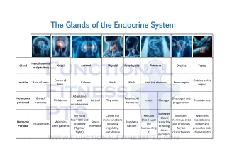
Common endocrine alterations in theelderly Menopause Estrogen (Progesterone?) Andro“pause” Testosterone DHT Somatopause GH Sarcopenia leanbody mass (appetite: CCK) Adreno“pause” DHEA DHEAS Cortisol ACTH FSH LH Failinglibido depression osteoporosis QOL issues Immuno-neuro-endocrine correlations Metabolic alterations Insulin resistance, IGT Metabolic syndrome (Carcinogenesis) “Synchropause” Melatonin Sleep(?) QOL issues certain autoimmune processes (inflammaging) Not “normal” ageing process, but common:subclinical hypo- and hyperthyroidism in elderly
Age-related endocrine alterations that promote frailty • Somatopause: decline in GH and IGF-I production leads to protein catabolism, loss of lean body mass, accumulation of fat • Adrenopause: decline in adrenal cortex-derived DHEA, DHEAS contribute to protein catabolism , loss of lean body mass and adiposity • Andro/Menopause: decline in sex steroids lead to protein catabolism, loss of lean body mass, diminished metabolic rate, accumulation of fat • Enhanced release and efficacy of peripheral anorexigenic, catabolic cholecystokininpromote insufficient food intake (e.g. protein deficiency) and loss of lean body mass • Insulin resistance and hyperinsulinemia induces deficient protein anabolism and enhanced fat accumulation
Factorsleadingtofrailty Aging Inflammatory state cardio-pulmonal hematological musculoskeletal neurological Genetic Frailty Hormonal changes Pathological changes Major musculoskeletal factors in frailty: Sarcopenia Osteoporosis Joint disease
The potential role of hormone replacement in the prevention of frailty • Numerous studies attest to the interactive influences between the GH/IGF-I and gonadal steroid axes. • Combined GH and sex steroid supplementation would increase or improve: • protein anabolism, skeletal muscle mass and strength; • decrease total and abdominal fat; • improve aerobic capacity and cardiovascular function; • various endocrine, metabolic, and other outcomes (e.g. sarcopenia, osteoporosis) • Dangerous side effects may include carcinogenesis, thrombosis, diabetes mellitus, etc.
Caloric restriction in the prevention of metabolic and endocrine complications • Caloric restriction decreases fat accumulation, insulin- and leptin-resistance, free radical production, inflammation. • Caloric restriction is the best way to delay both aging and cancer, affecting genes controlling these processes. • Geroprotector antioxidants (e.g. resveratrol) some hormones (DHEA), peptides and drugs are mimetics of calorie restriction to a certain degree. • Thus, caloric restriction presents an adequate and safe intervention for healthy aging and prevention of age-associated diseases, cancer included.
The spectrum of caloric intake from insufficient to excessive calories Longevity Cancer Autoimmune disease Oxidative stress Positive effects Regulated diet ↑ Calories Negative effects Hypothetical U-shaped curve over the spectrum of caloric intake from insufficient to excessive calories, emphasizing negative physiologic effects at both extremes and positive or hormetic effects within a range of restricted caloric intake. Energy deficit Energy excess Parenchymal cell number Loss of function Starvation Death Longevity Cancer Autoimmune disease Oxidative stress
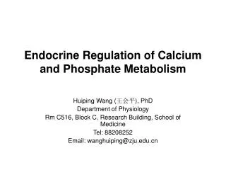
Age-related changes in glucose metabolism • Fasting glucose (FG) increases modestlybut significantly from early adulthood to the middle-aged years and then tends to remain constant. (Normal value: 3.8-6.0 mmol/L, impaired FG: 6.1-6.9 mmol/L, diabetes mellitus: from 7.0 mmol/L.) • In contrast, there is a striking increase in the 2-h glucose concentration during an oral glucose tolerance test (OGTT) throughout the adult years. In the course of OGTT the normal value for 2-h glucose concentration: below 7.8 mmol/L, IGT: 7.8-11.0 mmol/L, diabetes mellitus above 11.0 mmol/L. • The percentage of impaired glucose tolerance (IGT) and thus, the number of individuals with type 2 diabetes mellitus (relative insulin deficiency with hyperinsulinemia and progressive insulin resistance) increases markedly with age.
Glucose tolerance tests*in different age-groups 12 70 11 50 10 60 9 40 30 Blood sugar (mM) 8 20 7 6 Age (years) 5 0 90 0 20 60 120 150 Time (min) * 50 g glucosep.o.
Age-related changes in glucose metabolism • Gradual development of insulin resistance with age, particularly in obese (resistin and inflammatory cytokine production in abnormal fat tissue suppresses insulin effects) patients. • Suppression of cellular metabolism (neither oxygen, nor glucose can be utilized normally). • With muscle wasting and inactivity the insulin independent glucose utilization is limited. • Mild age-related hyperglycemia (insulin-resistance) is not to be treated by drugs. Treatment-associated hypoglycemia presents higher risk for the patient.
Complications of age-related disorders in carbohydrate metabolism • Acute complications include hyperglycemic hyperosmolar syndrome involving such cerebrovascular and metabolic mechanisms that lead to severe impairment of neuronal functions and even to coma with high mortality. • Long-term consequences include nonspecific complications, e.g. progressive atherosclerosis and hypertension (AMI, stroke, peripheral artery disease). • Specific long-term complications of DM affect the microcirculation (first enhancing dilution and permeability, later causing obstructions and tissue ischemia) and neurons. Chronic DM lead to the development diabetic retinopathy (blindness), diabetic nephropathy (from initial microalbuminuria to glomerulosclerosis, peritubular interstitial fibrosis, papilla necrosis and finally chronic renal failure) anddiabetic neuropathy. The latter involves microcirculatory damage of individual nerves or hyperglycemia-induced peripheral autonomic, sensory and motor neuropathy.
Prevention and treatment of age-related disorders in carbohydrate metabolism • Maintenance (or regain) of healthy BMI and body composition (prevention of adiposity) with help of low-calorie diet (avoiding refined sugars and fat) and physical activity (the latter reduces the need for insulin also via increasing insulin-independent glucose uptake of active muscles). • Inhibition of gastrointestinal glucose absorption • Inhibition of gluconeogenesis (biguanids) • Enhancement of insulin release (sulphanylureas) with side-effect of hyperinsulinemia and progressive burn-out of pancreatic beta-cells. • Enhancement of insulin sensitivity via stimulation of PPAR-receptors (thiazolidindiones with side-effect of adiposity, or angiotensine II receptor inhibitors, the metabolites of which also stimulates PPAR-g). • Stimulation of glucagon-like peptide-1 (GLP-1) and/or inhibition of its metabolism via dipeptidyl peptidase IV (DPP-4) inhibitors • Insulin(large doses)
Visceral obesity: In the course of aging prevalence of multimetabolic or metabolic X syndrome increases. Ectopic fat accumulation – lipotoxicity:Increased fat accumulation in various tissues (muscle, liver, myocardium, pancreas). Tissue functions deteriorate as a consequence: lower insulin efficacy, impaired contractility, diminished insulin release in response to elevated serum glucose level. Dyslipidemia – atherosclerosis: High serum cholesterol and dyslipidemia promote atherosclerosis. Diminished lipid utilization/fat metabolism due to impaired mitochondrial functions, lower energy consumption Prevention includes low-fat, low-calorie diet, physical activity, intake of omega-3 polyunsaturated fatty acids, administration of cholesterol synthesis inhibitor statins. Age-related changes in lipid metabolism
Gout • Definition • Gout is a disease involving precipitation of crystals of uric acid in tissues of the body. • Uric acid is a metabolite of purin break-down, a product of the enzyme xanthin-oxidase (XO). • Serum urate levels vary with age and sex. • Children: serum urate concentrations of 3.0-4.0 mg/dL(178-238 mmol/L). • Levels rise during puberty, remain low until menopause. • Adult men: 6.8 mg/dL (404 mmol/L) ,premenopausal women: 6 mg/dL (357 mmol/L) • In adulthood: concentrations rise steadily over time and vary with height, body weight, blood pressure, renal function and alcohol intake.
Hyperuricemia1 • Causes of increased se urate levels: • Primary hyperuricemia: the cause is innate • Secondary hyperuricemia: is the result of an acquired disorder. • 1 Increased production (40% of our urate production is of external, 60% is of internal origin) • 2 Decreased excretion
Hyperuricemia2 • Normal serum level of urate is 6.4 mg/dL (375 mmol/l). • At this level gout very rarely occurs (0.5%). • Above 7 mg/dl (416 mmol/L) the incidence of gout increases, above 9 mg/dL(535 mmol/L) therisk of gout is 90%. • The solubility of urate is decreased with cold and acidemia. → It is precipitated in the form of needle-like Na-urate crystals. Causes of increased se urate levels: • 1 Increased production • 2 Decreased excretion
1 Increased urate production External (food) sources of purines are • nuts (hazelnuts, peanuts, walnuts), • meat and liver (high content of cell nuclei). • Alcohol intake, overweight, hypertension, susceptibility to type 2 diabetes mellitus, male gender and aging → risk for gout. Internalurate production: • massive cell necrosis, cell proliferation • hypoxia (xanthine-dehydrogenase→ XO ) • alcohol ( XO), acidic metabolites →urate precipitation. • Defect of the salvage pathway
Evaluation of hyperuricemia • Hyperuricemia does not necessarily represent a disease, nor is it a specific indication for therapy. • The decision to treat depends on the cause and the potential consequences of the hyperuricemia in each individual.
2 Decreased urate excretion 90% of urate is excreted via the kidneys In the kidneys proximal tubuli reabsorb 97% of the urate, in the distal tubuliurate is secreted. Altogether 6-7% of the urate is eliminated. Secretion is antagonized by organic acids (lactate, ketone bodies – DM!, starvation; salicylate intoxication), alcohol, thiazide diuretics and may be impaired in kidney damage (lead poisoning), in genetic disorders or by stress. • 10% is excreted via the GI tract. Nicotinic acid administered to decrease serum lipid levels may block intestinal excretion.
Pathomechanism of acute consequences • Acute inflammation with fever, anorexia (the joint is swollen, red, with shiny skin and is extremely painful). • Role of neutrophil granulocytes in acute attacks of gout • In the background of symptoms futile activation of neutrophil granulocytes are presumed. • Following phagocytosis of Na-urate crystals, release of lysosomal enzymes, free radicals (oxidative burst) and inflammatory cytokines induce inflammation and tissue damage without destruction of the urate crystals. • Inhibition of neutrophil activation by colchicin efficiently suppresses acute symptoms. • Acute attacks are reversible within a few days without treatment. Without suppression of serum urate level they become recurrent.
Pathomechanism ofchroniccomplications • Chronic inflammation:fibrotic tissue and macrophagessurround the crystals→ foreign body granuloma = TOPHUS(inuntreated, neglectedcases). • Joints that are distal, therefore their temperature islower with pre-existing damage (mild arthrosis)facilitate the gouty arthritis. • PODAGRA: inflammation of the first metatarso-phalangeal joint (bigtoe). • Periodical acute recurrent episodes lead tothechronic phase, when joints are distorted, tophuses appear in the joints. Urateprecipitatesin the kidneys (urate stones, urate nephropathy) and in practically any tissue, except the brain (atherosclerosis, ischemic heart disease).
Other consequences Urate precipitation • kidneys • urolithiasis (hyperuricemia presents a 1,000× risk) • parenchymal damage (chronic sclerotising interstitial nephritis) • joints, leading to arthrosis • arterial wall - atherosclerosis. • coronaries - producing ischemic heart disease
Treatment of hyperuricemia • Diet A diet based on vegetables with little meat and nuts and no alcohol may help reduce hyperuricemia. • Xanthine-oxidase inhibitorAllopurinol reduces serum urate levels • Pain-killers Although salicylates tend to inhibit urate excretion as organic acids, in clinical practice they are used to suppress inflammation and pain in gout . • ColchicinDespite toxic side effects, efficacy in relieving gouty pain justifies its use.