Download
1 / 56
580 likes | 832 Views
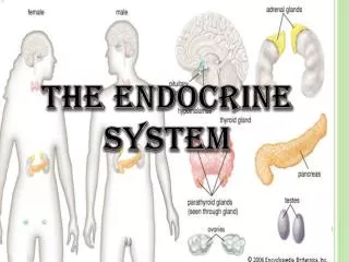
16. The Endocrine System: Part B. The Anatomy and Orientation of the Pituitary Gland. Figure 18.6a, b. Major Endocrine Organs: Pituitary (Hypophysis). Pituitary gland – two-lobed organ that secretes nine major hormones

E N D
16 The Endocrine System: Part B
The Anatomy and Orientation of the Pituitary Gland Figure 18.6a, b
Major Endocrine Organs: Pituitary (Hypophysis) • Pituitary gland – two-lobed organ that secretes nine major hormones • Neurohypophysis – posterior lobe (neural tissue) and the infundibulum • Receives, stores, and releases hormones from the hypothalamus • Adenohypophysis – anterior lobe, made up of glandular tissue • Synthesizes and secretes a number of hormones
Pituitary-Hypothalamic Relationships: Posterior Lobe • Is a down growth of hypothalamic neural tissue • Has a neural connection with the hypothalamus (hypothalamic-hypophyseal tract) • Nuclei of the hypothalamus synthesize oxytocin and antidiuretic hormone (ADH) • These hormones are transported to the posterior pituitary • Stores antidiuretic hormone (ADH) and oxytocin • ADH and oxytocin are released in response to nerve impulses • Both use PIP-calcium second-messenger mechanism at their targets
http://www.emc.maricopa.edu/faculty/farabee/BIOBK/BioBookENDOCR.html#Mechanisms%20of%20Hormone%20Actionhttp://www.emc.maricopa.edu/faculty/farabee/BIOBK/BioBookENDOCR.html#Mechanisms%20of%20Hormone%20Action
Neurohypophysis hormones Hormones that are produced in the hypothalamus and stored in the neurohypophysis
Oxytocin • Stimulates uterine contractions during childbirth by mobilizing Ca2+ through a PIP2-Ca2+ second-messenger system • Also triggers milk ejection in women producing milk • Plays a role in sexual arousal and orgasm in males and females
Antidiuretic Hormone (ADH) • Hypothalamic osmoreceptors respond to changes in the solute concentration of the blood • If solute concentration is high • Osmoreceptors depolarize and transmit impulses to hypothalamic neurons • ADH is synthesized and released, inhibiting urine formation • We will talk about this hormone in details with the urinary system
The anterior lobe • Is an outpocketing of the oral mucosa from epithelial tissue • There is no direct neural contact with the hypothalamus • Hormone production is regulated by the hypothalamus • Regulatory factors from the hypothalamus arrive directly to the adenohypophysis through the hypophyseal portal system • Subdivided into the • pars distalis – the largest and most anterior portion • pars intermedia – a narrow band between the pars distalis and the posterior lobe • pars tuberalis – wraps around the proximal portion of the infundibulum
Hypophyseal portal system • Portal system - a system of blood vessels that begins and ends in capillaries. The blood, after passing through one capillary bed, is passing through a second capillary network. • All blood entering the portal system will reach the target cells before returning to the general circulation
Pituitary-Hypothalamic Relationships: anterior Lobe • The hypophyseal portal system, consisting of: • The primary capillary plexus in the infundibulum • The hypophyseal portal veins • The secondary capillary plexus
Activity of the Adenohypophysis • The hypothalamus sends a chemical stimulus to the anterior pituitary • Releasing hormones stimulate the synthesis and release of hormones • Inhibiting hormones shut off the synthesis and release of hormones • The hormones of the anterior pituitary (7) are called tropic/trophic hormones because they “turn on” other glands or organs
Anterior Pituitary Hormones • All are proteins • All except GH activate cyclic AMP second-messenger systems at their targets • TSH, ACTH, FSH, and LH are all tropic hormones (regulate the secretory action of other endocrine glands)
Gonadotropins • Follicle-stimulating hormone (FSH) and luteinizing hormone (LH) • Secreted by gonadotrophs of the anterior pituitary • FSH stimulates gamete (egg or sperm) production • LH promotes production of gonadal hormones • Absent from the blood in prepubertal boys and girls • Regulation of gonadotropin release • Triggered by the gonadotropin-releasing hormone (GnRH) during and after puberty • Suppressed by gonadal hormones (feedback) • We will discuss these hormones later with the reproductive system
Prolactin (PRL) • Secreted by lactotrophs of the anterior pituitary • Stimulates milk production • Regulation of PRL release • Primarily controlled by prolactin-inhibiting hormone (PIH) • Blood levels rise toward the end of pregnancy • Suckling stimulates PRH release and promotes continued milk production
Growth Hormone (GH) or somatotropin • GH is an anabolic (tissue-building) hormone • Stimulate most body cells to increase in size and divide by increasing protein synthesis • Major target tissues are bone, cartilage and skeletal muscle • GH release is regulated by • Growth hormone–releasing hormone (GHRH) • Growth hormone–inhibiting hormone (GHIH) (somatostatin
Growth Hormone (GH) or somatotropin • The stimulation of growth by GH involves 2 mechanisms: • The primary one is indirect and more understood: • GH influence the liver, skeletal muscle, bone, and cartilage to release insulin-like growth factors (IGF)/somatomedins • The IGF binds to specific receptors on cells and increase the uptake of amino acids and their incorporation into new proteins
Growth Hormone (GH) or somatotropin • Direct effects • In ET and CT stimulate cell division and differentiation (the subsequent cell growth is mediated by IGF) • In adipose tissue GH stimulates the breakdown of stored triglycerides by adipocytes and the release of fatty acids to the blood. That promotes the use of fatty acid for energy instead of the use of glucose (glucose-sparing effect)
Thyroid-Stimulating Hormone (Thyrotropin) • Produced by thyrotrophs of the anterior pituitary • Stimulates the normal development and secretory activity of the thyroid • Regulation of TSH release • Stimulated by thyrotropin-releasing hormone (TRH) • Inhibited by rising blood levels of thyroid hormones that act on the pituitary and hypothalamus
Adrenocorticotropic Hormone (Corticotropin) • Secreted by corticotrophs of the anterior pituitary • Stimulates the adrenal cortex to release corticosteroids • Regulation of ACTH release • Triggered by hypothalamic corticotropin-releasing hormone (CRH) in a daily rhythm • Internal and external factors such as fever, hypoglycemia, and stressors can alter the release of CRH
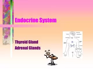
Thyroid Gland The thyroid gland on the anterior side of the neck. The thyroid gland has a right lobe and a left lobe connected by a narrow isthmus http://webanatomy.net/histology/endocrine_histology.htm
Thyroid gland histology • The thyroid gland contains thyroid follicles lined with simple cuboidal epithelium – follicular cells • The follicle cells surround a follicular cavity that contain the colloid; a fluid that contains a large amount of proteins - thyroglobulin that contain the amino acid tyrosine • Each follicle is surrounded by a capillary network. • Between the follicles C cells/parafollicular cells can be found
Thyroid Hormone • Thyroid hormone – major metabolic hormone • Consists of two related iodine-containing compounds • T4 – thyroxine; has two tyrosine molecules plus four bound iodine atoms • T3 – triiodothyronine; has two tyrosines with three bound iodine atoms
Synthesis of Thyroid Hormone • Thyroglobulin is synthesized by the follicular cells and released into the lumen • Iodides (I–) are actively taken into the cell by membrane carrier proteins • The iodide ions diffuse to the apical surface of the cells (these cells are facing towards…?), oxidized to iodine (I2) by the enzyme thyroid peroxidase and released to the colloid. • Iodine attaches to tyrosine in the thyrogobulin, forming T1 (monoiodotyrosine, or MIT), and T2 (diiodotyrosine, or DIT)
Synthesis of Thyroid Hormone • Iodinated tyrosines link together to form T3 and T4 • Coupling reaction MIT + DIT T3 / triiodothyronine DIT + DIT T4 / thyroxin (tetraiodothyronine) • The colloid is then endocytosed and combined with a lysosome, where T3 (10%) and T4 (90%) are cleaved and diffuse into the bloodstream • 75% of the T4 and 70% of the T3 are transported attached to thyroid-binding protein (TBGs) and the rest to a special albumin
Thyroid Hormone and target cells • Thyroid hormones influence almost every cell of the body • Inside the cells they bind to receptors in one of 3 locations: • In the cytoplasm – storage of thyroid hormones to be released if the intracellular levels decrease • On the mitochondria surface – increase rate of ATP production • In the nucleus – activate genes that control the synthesis of enzymes that involve with energy production and utilization (for example increase of production of sodium-potassim ATPase that uses ATP)
Other effects of Thyroid Hormone • TH is concerned with: • Activate genes that code for enzymes that are involved in glycolysis (Glucose oxidation) • Increasing metabolic rate of the cells • Increase heat production (calorigenic effect) • Although the major thyroid hormone that is being produced is the T4 (90%) T3 is the one responsible for the TH effects • Enzymes in the kidneys, liver and other tissues convert T4 to T3
Transport and Regulation of TH • Negative feedback regulation of TH release • Rising TH levels provide negative feedback inhibition on release of TSH • Hypothalamic thyrotropin-releasing hormone (TRH) can overcome the negative feedback during pregnancy or exposure to cold
Calcitonin • A peptide hormone produced by the parafollicular, or C cells • Lowers blood calcium levels • Antagonist to parathyroid hormone (PTH)
Calcitonin • Calcitonin targets the skeleton, where it: • Inhibits osteoclast activity (and thus bone resorption) and release of calcium from the bone matrix • Stimulates calcium uptake and incorporation into the bone matrix • Regulated by a humoral (calcium ion concentration in the blood) negative feedback mechanism
Parathyroid Glands • Four to eight tiny glands embedded in the posterior aspect of the thyroid • Contain oxyphil cells (function unknown) and chief cells that secrete parathyroid hormone (PTH) or parathormone • PTH—most important hormone in Ca2+ homeostasis
Effects of Parathyroid Hormone • PTH release increases Ca2+ in the blood as it: • Stimulates osteoclasts to digest bone matrix • Enhances the reabsorption of Ca2+ and the secretion of phosphate by the kidneys • Increases absorption of Ca2+ by intestinal mucosal • Rising Ca2+ in the blood inhibits PTH release
Adrenal (Suprarenal) Glands • Adrenal glands – paired, pyramid-shaped organs atop the kidneys • Structurally and functionally, they are two glands in one • Adrenal medulla – neural tissue; part of the sympathetic nervous system • Adrenal cortex - three layers of glandular tissue that synthesize and secrete corticosteroids
http://www.histology-world.com/photomicrographs/adrenallabel.jpghttp://www.histology-world.com/photomicrographs/adrenallabel.jpg
Adrenal Cortex • Synthesizes and releases steroid hormones called corticosteroids • Different corticosteroids are produced in each of the three layers • Zona glomerulosa – glumerulus- little ball. Secretes mineralocorticoids – main one aldosterone • Zona fasciculata – glucocorticoids (chiefly cortisol) • Zona reticularis – gonadocorticoids (chiefly androgens)
Zona glumerulosa - Mineralocorticoids • Aldosterone secretion is stimulated by: • Rising blood levels of K+ • Low blood Na+ • Decreasing blood volume or pressure
Zona glumerulosa - Mineralocorticoids • The mineralocorticoids are steroids that affect the electrolytes composition of the body extracellular fluids. • Aldosterone – most important mineralocorticoid • Maintains Na+ balance by reducing excretion of sodium from the body • Stimulates re-absorption of Na+ by the kidneys • Prevents the loss of Na+ by the kidneys, sweat glands, salivary glands and digestive system • As a result of Na+ reabsorption there is also water reabsorption • The retention of Na+ is accompanied by a loss of K+ • These effects will be discussed later (urinary system) • The effect of aldosterone is the most effective when normal ADH levels are present.
Zona fasciculata - Glucocorticoids (Cortisol/hydrocortisone) • This adrenal layer responds to ACTH (which endocrine glands secretes ACTH?) • Main hormone secreted are the Cortisol/hydrocortisone and small amounts of corticosterone • Glucocorticoids accelerate the rates of glucose synthesis and glycogen formation – especially in the liver • Adipose tissue responds by releasing fatty acids into the blood and the tissues start to utilize fatty acids as source of energy - glucose-sparing effect (which other hormone as similar effect?) • Clucocorticoids also have anti-inflammatory effect – inhibit the activities of WBC (use?)
Zona reticularis Gonadocorticoids (Sex Hormones) • Most gonadocorticoids secreted are androgens (male sex hormones), and the most important one is testosterone • Androgens can be converted into estrogens after menopause • Both hormones from the kidney origin do not effect sexual characteristics
Adrenal Medulla • Secrete epinephrine and norepinephrine • Epinephrine is the more potent stimulator of the heart and metabolic activities • Norepinephrine is more influential on peripheral vasoconstriction and blood pressure
Epinephrine and Norepinephrine • increase in the rate and strength of the heartbeat resulting in increased blood pressure; • Increase skeletal muscle strength and endurance by increasing glucose breakdown • Increases availability of fatty acids from adipose tissue • Induce blood shunted from the skin and viscera to the skeletal muscles, coronary arteries, liver, and brain; • Cause rise in blood sugar; • Trigger bronchi to dilate to assists in pulmonary ventilation; • pupils dilate; • Reduce clotting time of the blood; • increased ACTH secretion from the anterior lobe of the pituitary. • All of these effects prepare the body to take immediate action
Pancreas • A triangular gland, which has both exocrine and endocrine cells, located behind the stomach • Acinar cells produce an enzyme-rich juice used for digestion (exocrine product) • Pancreatic islets (islets of Langerhans) produce hormones (endocrine products)
Pancreas – islets of Langerhans cells • The islets contain two major cell types: • Alpha () cells that produce glucagon • Beta () cells that produce insulin • The islets also contain • Delta cells – produce a peptide hormone identical to GH inhibiting hormone (GH-IH). That hormone supresses the release of glucagon and insulin and slows food absorptopn and digestive enzyme secretion • F cells – Produce the hormone pancreatic polypeptide (pp) that inhibits gallbladder contractions and regulate the production of some pancreatic enzymes
Glucagon • Major target is the liver, where it promotes • Glycogenolysis—breakdown of glycogen to glucose • Gluconeogenesis—synthesis of glucose from lactic acid and noncarbohydrates • Release of glucose to the blood
Glucagon • A 29-amino-acid polypeptide hormone that is a potent hyperglycemic agent • Its major target is the liver, where it promotes: • Glycogenolysis – the breakdown of glycogen to glucose • Gluconeogenesis – synthesis of glucose from lactic acid and noncarbohydrates • Release of glucose to the blood from liver cells
Insulin • A 51-amino-acid protein consisting of two amino acid chains linked by disulfide bonds • Insulin is released when glucose levels exceed normal levels (70-110 mg/dl) http://www.chemistryexplained.com/images/chfa_02_img0437.jpg
Effects of Insulin Binding • Insulin facilitates entry of glucose cells by binding to a membrane receptor • The complex insulin-receptor make a specific carrier protein (GLUT4) available • Once at the cell surface, GLUT4 facilitates the passive diffusion of circulating glucose down its concentration gradient into cells. • Receptors for insulin are present in most cell membranes (insulin-dependant cells) • Cells that lack insulin receptors are cells in the brain, kidneys, lining of the digestive tract and RBC (insulin-independent cells). • Those cells can absorb and utilize glucose without insulin stimulation.
Effects of Insulin Binding • Insulin effects: • Acceleration of glucose uptake as a result from an increase of the number of glucose carrier proteins • Acceleration of glucose utilization and increased ATP production • Stimulation of glycogen formation in the liver and muscle cells • Inhibits glycogenolysis (break down of glycogen) and gluconeogenesis (glucose building) • Stimulation of amino acid absorption and protein synthesis • Stimulation of triglyceride formation in adipose tissue • As a result glucose concentration in the blood decreases