Download
1 / 35
350 likes | 362 Views
Cardiovascular System. Heart – cardiac muscle physiology Cardiac conduction system Heart structures Vessels- arteries and veins Blood – WBC, RBC and Platelets, =Formed elements plus the plasma (the fluid). Types of Cardiac Cells.

E N D
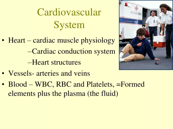
Cardiovascular System • Heart – cardiac muscle physiology • Cardiac conduction system • Heart structures • Vessels- arteries and veins • Blood – WBC, RBC and Platelets, =Formed elements plus the plasma (the fluid)
Types of Cardiac Cells • Auto-rhythmic cells = pacemaker cells (depolarize spontaneously) • Contractile cells – myofibrils –connected well together to form “functional Syncitium” for myocardium contraction Ease of movement between cells (conductivity)
Mechanical and Electrical • Critical to remember that there is mechanical aspects to heart function=myofibrils and electrical aspect • Pacemaker cells and • Resulting AP from these cells to working muscle cells causes depolarization and repolarization
Autorhythmic Cells Cells fire spontaneously, act as pacemaker and form conduction system for the heart SA node cluster of cells in wall of Rt. Atria begins heart activity that spreads to both atria excitation spreads to AV node AV node in atrial septum, transmits signal to bundle of His AV bundle of His the connection between atria and ventricles Bundle branches To right and left side of heart along ventricular septum Purkinje fibers, large diameter fibers that conduct signals quickly in ventricles Conduction System of Heart
Rhythm of Conduction System • SA node fires spontaneously 80-100 times per minute • AV node fires at 40-60 times per minute • If both nodes are suppressed fibers in ventricles by themselves fire only 20-40 times per minute • Artificial pacemaker needed if pace is too slow • Extra beats forming at other sites are called ectopic pacemakers • caffeine & nicotine increase activity
Timing of Atrial & Ventricular Excitation • SA node setting pace since is the fastest • In 50 msec excitation spreads through both atria and down to AV node • 100 msec delay at AV node due to smaller diameter fibers- allows atria to fully contract filling ventricles before ventricles contract • In 50 msec excitation spreads through both ventricles simultaneously
Physiology of Contraction • Depolarization, plateau, repolarization
Important Electrolytes • Sodium – major extracellular ion • Potassium - major intracellular ion • Chloride - negative ion • Calcium - very important for muscle contraction
Normal Resting Membrane Outside cell Inside cell
Protein Channels • Special channels allow cations to go through membrane Will cause membrane charge to rise towards 0mV
Action Potential Origin • Channel proteins • Voltage gated ion channels • potassium • sodium
Sodium & Potassium Channels • Responds to concentration gradients • “depolarization” = movement away from resting potential towards or above 0 mV • “repolarization” = return to resting potential -90 mV • Necessity of channels
Overview of Ion Movement Inside cell
Depolarization • Action potential movement = ion exchange • Sodium moves into cell • Potassium moves out of cell • Membrane potential rises 0 -70
this picture shows a neuron • the principle is the same in a cardiac cell
Sodium and Potassium ions utilize both fast and slow channels Sodium enters cells via slow channels until “threshold” is reached Mass of sodium then enters cell via the fast channels during contraction Fast and Slow Channels
Repolarization • Bringing the membrane back to its resting state • Active pump moves ions against the gradients • Sodium/Potassium Pump
Sodium/Potassium Pump • Maintains concentration • ~ 80% of metabolic energy consumed by pump • “Leaky membrane” • Sodium leaks in • Potassium leaks out
Phases of the Action Potential • Specific sequence of events • Phases 0 through 4 • Important to know what happens in each phase • Comprehension of pathology and pharmacotherapy
Phases of Action Potential • Phase 0 = Na+ channels open slow then fast • Phase 1 = K+ channels open slow
Phases of Action Potential • Phase 2 = Ca+ channels open • Phase 3 = K+ channels open
Phases of Action Potential • Phase 4 - Na/K pump returns membrane to rest • Na+ leaks = • Slow phase 4 rise = • automaticity
The Condensed Version • Maintenance of membranes resting potential • External force (stimulus) • Slow rise brings it to threshold at - 70 mV • Ion exchange begins • Na+, then Ca+ into cell • K+ out of cell
Depolarization & Repolarization • Depolarization • Cardiac cell resting membrane potential is -90mv • excitation spreads through gap junctions • fast Na+ channels open for rapid depolarization • Plateau phase • 250 msec (only 1msec in neuron) • slow Ca+2 channels open, let Ca +2 enter from outside cell and from storage in sarcoplasmic reticulum, while K+ channels close • Ca +2 binds to troponin to allow for actin-myosin cross-bridge formation & tension development • Repolarization • Ca+2 channels close and K+ channels open & -90mv is restored as potassium entering the cell • Refractory period • very long so heart can fill with blood
Action Potential in Cardiac Muscle Changes in cell membrane permeability.
Electrocardiogram---ECG or EKG • EKG • Action potentials of all active cells can be detected and recorded • P wave • atrial depolarization • P to R interval • conduction time from atrial to ventricular excitation • QRS complex • ventricular depolarization • T wave • ventricular repolarization
One Cardiac Cycle • At 75 beats/min, one cycle requires 0.8 sec. • systole (contraction) and diastole (relaxation) of both atria, plus the systole and diastole of both ventricles • Stroke volume (SV) • the volume ejected per beat from each ventricle, about 70ml MEDIC Point: What would the normal amount of blood pumped out of the Left ventricle be in 1 minute?
Phases ofCardiac Cycle • Isovolumetric relaxation • brief period when volume in ventricles does not change--as ventricles relax, pressure drops and AV valves open • Ventricular filling • rapid ventricular filling:as blood flows from full atria • diastasis: as blood flows from atria in smaller volume • atrial systole pushes final 20-25 ml blood into ventricle • Ventricular systole • ventricular systole • isovolumetric contraction • brief period, AV valves close before SL valves open • ventricular ejection: as SL valves open and blood is ejected
Ventricular Pressures • Blood pressure in aorta is 120mm Hg • Blood pressure in pulmonary trunk is 30mm Hg • Differences in ventricle wall thickness allows heart to push the same amount of blood with more force from the left ventricle • The volume of blood ejected from each ventricle is 70ml (stroke volume) • Why do both stroke volumes need to be same?