Download
1 / 36
370 likes | 532 Views
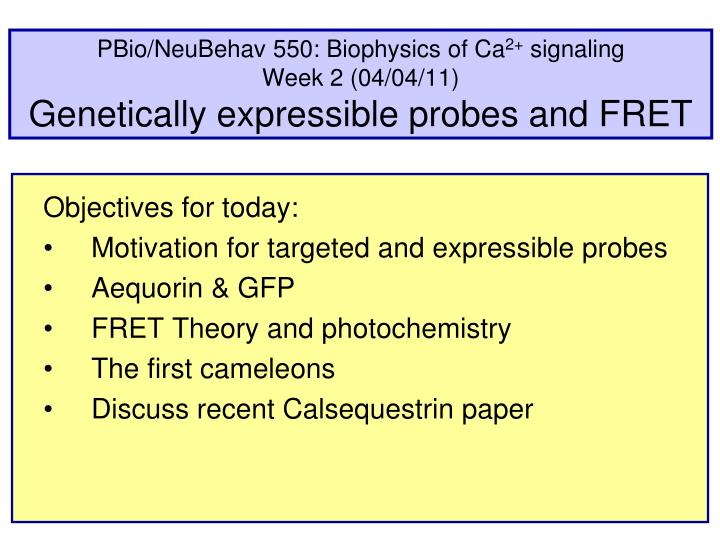
PBio/NeuBehav 550: Biophysics of Ca 2+ signaling Week 2 (04/04/11) Genetically expressible probes and FRET. Objectives for today: Motivation for targeted and expressible probes Aequorin & GFP FRET Theory and photochemistry The first cameleons Discuss recent Calsequestrin paper.

E N D
PBio/NeuBehav 550: Biophysics of Ca2+ signalingWeek 2 (04/04/11)Genetically expressible probes and FRET Objectives for today: Motivation for targeted and expressible probes Aequorin & GFP FRET Theory and photochemistry The first cameleons Discuss recent Calsequestrin paper
Standard tools for calcium studies Tools for calcium studies [Caged calcium] [NP-EGTA] [–—NP] The original Ca/Mg chelator & buffer EDTA (1946) EGTA (1955) BAPTA (1980) Fura, Indo Ca Green Ca-selective chelator & buffer slow, pH sensitive Roger Tsien’s fast buffers & fluorescent indicators KCa ~ 80-300 nM
Proteins as indicators Advantages of proteins as indicators Binding site already evolved for high selectivity Can be further engineered by directed or random mutation Genetically expressible by transfection, viral infection, transgenic lines Targetable to specific cell types at specific times in organisms use promoters and enhancers Targetable to subcellular locations and organelles in cells add nuclear, mitochondrial, ER, etc. targeting sequences No need for pipettes or tricky loading procedures Do not leak out of cell or organelle Some are more resistant to bleach than conventional dyes
Aequoria Fluorescent proteins make Aequorea glow at 508 nm Green fluorescent ring ---Shimomura O, Johnson FH, Saiga Y, 1962, Extraction, purification and properties of Aequorin, a biolumi-nescent protein from the luminous hydromedusan, Aequorea. J. Cell. Comp. Physiol., 59: 223-239. [470 nm] ---R.Y. Tsien, 1998, The Green FluorescentProtein, Annual Review of Biochemistry 67, pp 509-544. [508 nm] Aequorea victoria from Puget Sound in brightfield and false color
Aequorin 2 Aequorin: a bioluminescent Ca2+ binding protein complex containing coelenterazine coelenterazine M.W. = 22,514 with four E/F hands Aequorin (Aeq) falls in the general heading of "luciferases" that bind a "luciferin" and luminesce in response to a ligand. (The most famous of these is firefly luciferase that can be used to measure ATP concentrations.) Reaction: Aeq + coelenterazine ----> Aeq.c [non-covalent complex] Aeq.c + ~3 Ca2+ ----> Ca3.Aeq.c* + CO2 Ca3.Aeq.c* -----> Ca3.Aeq.c** + [blue photon--470 nm] Aequorin is therefore a one-shot calcium detector with a non-linear Ca2+ dependence of luminescence. It is "consumed" by a detection event.
Ca2+ fluxes in an excitable cell Typical Ca2+ fluxes in a non-excitable cell Inputs: hormones, cytokines, growth factors, antigens PIP2 Agonist Na+-Ca2+ exchanger R DAG Gq PLC IP3 LDCSG Ca2+ Ca2+ Ca2+ IP3R channel Na+ SERCA pump ATP ER Plasma membrane Ca2+ ATP Ca2+ Mito PM Ca2+ ATPase SOC/CRAC channel Ca2+ Na+ Responses: Fluid secretion, exocytosis, channel gating, enzyme activities, cell division, proliferation, gene expression
Biological example aequorin Aequorin reports [Ca] outside and inside mitochondria FCCP will depolarize inner membrane of mitochondrion 10 histamine stimulus Mito Aeq targeted inside mitochondrial matrix [Ca] (mM) Aeq targeted between mitochondrial membranes Control: Ca sucked into mitochondrion 5 histamine stimulus 4 Control + 5 mM FCCP: Ca does not enter [Ca] (mM) + 5 mM FCCP 0 time Two batches of HeLa cells transfected with different aequorin constructs. One targets to the inter-membrane space of mitochondria and the other targets all the way to the matrix of the mitochondria. Cells were then soaked in micromolar coelenterazine at zero calcium for several hours. (Rizzuto...Pozzan, Science, 1998)
napthalene anthracene tetracene absorption spectra small box, short wave large box, long wave Spectra and particle-in-a-box (think organ pipes) coelenterazine emits 470 nm Tyrosine/ phenol: Exc. 275 nm, emits 310 nm)
GFP GFP: generates a fluorescent chromophore from its amino acids autocatalytically Maturation can be slow Engineer codons folding color photoconversion Note: N- and C- are close to each other M.W. = 26,938 dehydration N C GFP, a beta barrel
Ph sensitive quantum yield of FPs pKa = 6.6 Dark form Habuchi S, Tsutsui H, Kochaniak AB, Miyawaki A, van Oijen AM. mKikGR, a monomeric photoswitchable fluorescent protein. PLoS ONE. 2008;3:e3944.
Photoswitchable FPs pKa = 6.6 Dark form Astrocytes mKikGR-actin 405 nm after 405 nm Habuchi S, Tsutsui H, Kochaniak AB, Miyawaki A, van Oijen AM. mKikGR, a monomeric photoswitchable fluorescent protein. PLoS ONE. 2008;3:e3944.
Förster/Fluorescence resonance energy transfer(FRET): A proximity detector (molecular ruler) that changes color FRET illustrate 440 nm 480 nm YFP hn CFP emission hn Separated: no FRET excitation no 440 nm excitation no hn 440 nm hn hn YFP FRET! CFP 535 nm Close together: FRET excitation emission Green fluorescent protein (GFP) has been engineered to make forms with various fluorescent colors (GFP, CFP, YFP, …). They have overlapping spectra and can transfer excitation directly by FRET when the proteins are close together. The energy transfer occurs without a photon.
Forster Eq FRET depends steeply on distance. R depends on overlap. Donor Acceptor 440 nm YFP FRET! CFP 535 nm excitation emission r fDeA Ro6 Ro6 + r 6 Transfer efficiency E: E = ------------- Förster formula for Förster radius Ro Ro = Const. {fdonk2J n –4} 1/6 Where fdon quantum efficiency of donor k orientation factor (0 – 4) n local refractive index J "overlap integral" of donor fluorescence (fD) and acceptor absorption eA J = 500 600 l = wavelength
More steps in the Jablonski diagram internal conversion (1 ps) (polar) solvent relaxation (100 ps) competition for re-radiation, quench, FRET, or other non- radiative (3 ns) absorption (1 fs) knr hnFRET fluorescence quench FRET Donor Acceptor
FRET as a ‘Spectroscopic Ruler’ The efficiency of energy transfer is proportional to the inverse of the sixth power of the distance separating the donor and acceptor fluorophore ECFP/EYFP Förster distance 30 Å Förster distance 50 Å e.g., ECFP/EYFP Förster distance 70 Å E % decreases as distance between donor and acceptor increases When two fluorophores separated by Förster distance (where r = Ro), E transfer is 50%
A family of Ca2+-sensitive switches and buffers Calmodulin helix-loop-helix makes E-F hand { x x x x KCa ~ 14 mM for free calmodulin Calmodulin MW ~ 17 kDa Calmodulin (CaM) : An abundant 149 amino acid, highly conserved cyto-plasmic protein with 4 binding sites for Ca2+ each formed by "EF-hands." Many other homologous Ca2+ binding proteins of this large EF-hand family act as Ca switches and Ca buffers. The Ca2+ ions bind cooperatively and become encircled by oxygen dipoles and negative charge. CaM com-plexes with many proteins, imparting Ca2+-dependence to their activities.
Calmodulin folds around a target helix Calmodulin folds How many Ca need to bind? MLCK peptide 4 Ca CaM Binding of Ca2+ to CaM causes CaM to change conformation. Binding of CaM to targets can increase the Ca2+ binding affinity of CaM greatly. The target peptide in this crystal structure is the regulatory domain of smooth-muscle myosin light-chain kinase (MLCK). The interaction of CaM and MLCK allows smooth muscle contraction to be activated in a Ca2+-dependent manner. (Meador WE, Means AR & Quiocho, 1992.)
Design of CaMeleons: Expressible proteins for Ca detection Design of CaMeleons: 440 nm 480 nm YFP Low calcium: No FRET C N CaM MLCK CFP C YFP 440 nm FRET CFP High calcium: FRET 535 nm N Two GFPs in one peptide interact by fluorescence resonance energy transfer (FRET). Targeting sequences can be added to direct constructs to specific compartments. (Miyawaki, Roger Tsien et al., 1997)
Genetic tailoring of first-generation cameleons Targeting CaM-M13 Cameleon name: N C 2 EGFP nls EGFP 2nu CRsig 3er EGFP KDEL EYFP YC2 CRsig EYFP KDEL YC3er CRsig EYFP YC4er KDEL Abbreviations: CRsig = calreticulin signal sequence nls = nuclear localization signal KDEL = ER retention signal (Miyawaki et al. and Tsien, Nature, 1997)
Localization Targeting of cameleons YC2 GC2nu YC3er scales = "10 mm"
How many calciums bind? Mutating calcium binding. green cameleon 1 fluorescence ratios 100 N C GC1 % of 510/445 nm emission ratio GC1/E31Q GC1/E104Q free calcium (M) Calcium binding and the conformation change can be tailored by making mutations in the EF hand regions of the calmodulin. Glutamate E31 is in the first EF hand (at p12') and E104 is in the third EF hand (also at p12').
Mutating calcium binding for ER/SR sensitivity YC4.3 E31Q YC2.1 D1 YC3.3 E104Q ER/SR Palmer AE, Jin C, Reed JC, Tsien RY. PNAS 2004
Canato et al. 2010 paperMassive alterations of sarcoplasmic reticulum freecalcium in skeletal muscle fibers lacking calsequestrinrevealed by a genetically encoded probe Sarah Nowakowski: Explain Question that paper investigates (not what it concludes). Explain SR and CSQ. No need for audience inclusion. Curtis Easton: Fig. 1A&B Merle Gilbert: Fig. 1C Aaron Williams: Fig. 2 Wucheng Tao: Fig. 3 Jennifer Deem: Fig. 4 Bertil: Fig. 5 --5 min per fig --give it a title --explain axes --ask leading questions to get students to discuss
Sarah Nowakowski: Explain Question that paper investigates (not what it concludes). Explain SR and CSQ. No need for audience inclusion. Massive alterations of SR free calcium in skeletal muscle fibers lacking calsequestrin revealed by a genetically encoded probe
CASQ2 gene expression in Allen Brain Atlas Cerebellar Pukinje neurons (mouse atlas) (mouse CASQ2 ISH)
Fig 1 A&B; Curtis Easton (A) Localization of D1ER on both sides of Z lines stained with α-actinin antibody (D1ER, green; α-actinin antibody + rhodamine, red). D1ER -actinin Canato M et al. PNAS 2010;107:22326-22331
Fig 1 C; Merle Gilbert (A) Localization of D1ER on both sides of Z lines stained with α-actinin antibody (D1ER, green; α-actinin antibody + rhodamine, red). D1ER Canato M et al. PNAS 2010;107:22326-22331
WT CSQ-KO Typical recordings of changes in intraluminal SR Ca2+ concentrations during repetitive stimulation at different stimulation rates. Fig 2 A, B, C; Aaron Williams YFP CFP 1 Hz 5 Hz 20 Hz Canato M et al. PNAS 2010;107:22326-22331
Fig 3; Wucheng Tao Average amplitudes of the decline in the YFP/CFP R (ΔR) during contractile activity with increasing stimulation frequency. Canato M et al. PNAS 2010;107:22326-22331
Fig 4; Jennifer Deem YFP/CFP Time course of the change in the YFP/CFP R during the SR refilling. YFP both are CSQ-KO fibers YFP/CFP YFP Canato M et al. PNAS 2010;107:22326-22331
Fig 5; Bertil WT CSQ-DKO Fibers lacking CSQ are not able to maintain a high cytosolic calcium concentration during repetitive stimulations. Fura-2 Fura-2 20-Hz stimulation Canato M et al. PNAS 2010;107:22326-22331
Cameleon emission combines two spectra EYFP ECFP Ca no Ca YC3.1 cameleon emission intensity emission ECFP EYFP There is FRET even with no Ca2+! Amount of FRET gives distances: ~5.0 and 6.5 nm, or 50 and 65 Å. This is not a large change.
Absorption and fluorescence spectra reflect internal energy levels Absorption bands S1 S1 Energy S0 S0 Jablonski diagram Absorption wavelength Absorber has several electronic states (S0, S1, S2, etc.). It also has vibrational states whose close spacing means that photons of a range of close energies can be absorbed. If the absorption spectrum has a second peak (at shorter wavelength), it is for excitation to S2 or because the dye has several molecular forms/conformations.
Lifetime & FRET FRET speeds donor and slows acceptor F Ca2+-bound CaMeleon 530 nm from EYFP by FRET emission intensity 480 nm from ECFP 0 2 4 6 time (ns) Fluorescence decays recorded with YC3.1 cameleon dissolved in buffer. Excitation at 420 nm excites the ECFP part. (Habuchi et al. Biophys J, 2002)
R5 R2' R7' R6 Electron withdrawing on Fluo dye series