Download
1 / 16
160 likes | 367 Views
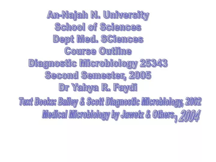
An-Najah N. University School of Sciences Dept Med. SCiences Course Outline Diagnostic Microbiology 25343 Second Semester, 2005 Dr Yahya R. Faydi. Text Books: Bailey & Scott Diagnostic Microbiology, 2002 Medical Microbiology by Jawetz & Others. 2004. ,. Aim of the Course.

E N D
An-Najah N. University School of Sciences Dept Med. SCiences Course Outline Diagnostic Microbiology 25343 Second Semester, 2005 Dr Yahya R. Faydi Text Books: Bailey & Scott Diagnostic Microbiology, 2002 Medical Microbiology by Jawetz & Others 2004 ,
Aim of the Course This course is a practical application of Medical Microbiology 25342 given in the first semester. The goals of the course are: 1- Study the different groups of microorganisms with emphasis on laboratory diagnosis. 2- Organ system infections with emphasis on specimen processing if needed, culture on the specific media & incubation under the suitable laboratory environment according to the type of the microorganism. Other methods of diagnosis such as microscopy, serology, molecular methods, chromatography. 3- Clinical specimens collection & transport. 4- The duty of the microbiologist towards the treating team & patients. 5- Laboratory design & instruments needed for a medical microbiology laboratory. Quality control for the different tests performed in the laboratory .
Functions of the microbiologist 1- Cultivation, identification & antimicrobial susceptibility testing of microorganisms. 2- Direct detection of infecting microorganisms by microscopy. 3- Direct detection of specific products of infecting organisms using chemical, immunologic or molecular techniques. 4- Detection of antibodies produced by the patient in response to an infecting organism (serodiagnosis). Role of the microbiologist: Responsibility to the patient & clinician Communication of laboratory findings Expediting results reporting-computerization General concepts for specimen collection & handling 1- Appropriate collection techniques. 2- Specimen transport. 3- Specimen preservation. 4- Specimen storage 5- Specimen labeling. 6- Specimen requisition.
7- Rejection of unacceptable specimen Specimen processing 1- gross examination of specimen. 2- Direct microscopic examination. 3- Selection of culture media. 4- Specimen preparation. 5- Inoculation of solid media. 6- Incubation conditions. 7- Extent of identification required.
Beta hemolytic Alpha haemolytic streptococci
Methods for Bacterial Identification Bacterial identification is central to diagnostic bacteriology because: 1- Determining the significance of the isolated pathogen ( pathogen or contaminant ). 2- Guiding physician care of the patient. 3- Determining whether laboratory testing for detection of antimicrobial resistance is warranted. 4- Determining the type of antimicrobial therapy that is appropriate. 5- Determining whether the pathogen is a risk to other people. Methods 1- Laboratory cultivation, isolation and identification of bacteria using macroscopic (colony) morphology, microscopic morphology (light, dark field, phase contrast, fluorescent, EM ) & staining characteristics ( gram stain & acid fast stain ), biochemical, serological, chromatography and molecular methods. 2- Molecular methods There are limitations for the previous methods as follows: a- Inability to grow certain fastidious pathogens. B- Inability to maintain viability during transport to the laboratory. C- Delay in cultivation & identification of certain pathogens ( Mycobacterium tuberculosis ). D- Lack of reliable methods for identification of certain pathogens.
The molecular methods include: 1- Nucleic acid hybridization methods It requires preparation of one nucleic acid strand (probe), DNA or RNA from known organism labeled with reporter which may be radioactiveP32, I125, S32,or biotin- avidin, digoxigenin or chemiluminescent labels.The target will be nucleic acid strand from the unknown organism. Thus the basic steps in hybridization assay include: 1- Preparation of labeled single - stranded probe nucleic acid. 2- Preparation of labeled single - stranded target nucleic acid. 3- Mixture & hybridization of target & probe. 4- Detection of hybridization by the reporter. 2- Amplification methods – Target Nucleic Acid Amplification – PCR Hybridization methods is not sensitive for clinical specimens containing target nucleic acid in very small amounts. This result in false negative results. Thus polymerase chain reaction (PCR) is needed for amplification of target followed by identification. The PCR involves 30 – 50 cycles, with each cycle comprising three sequential reactions: denaturation of target nucleic acid, primer annealing to single-stranded target nucleic acid, & extension of primer target duplex.
The purpose of a PCR (Polymerase Chain Reaction) is to make a huge number of copies of a gene. This is necessary to have enough starting template for sequencing. • The cycling reactions : • There are three major steps in a PCR, which are repeated for 30 or 40 cycles. This is done on an automated cycler, which can heat and cool the tubes with the reaction mixture in a very short time. • Denaturation at 94°C : During the denaturation, the double strand melts open to single stranded DNA, all enzymatic reactions stop (for example : the extension from a previous cycle). • Annealing at 54°C :The primers are jiggling around, caused by the Brownian motion. Hydrogen bonds are constantly formed and broken between the single stranded primer and the single stranded template. The more stable bonds last a little bit longer (primers that fit exactly) and on that little piece of double stranded DNA (template and primer), the polymerase can attach and starts copying the template. Once there are a few bases built in, the hydrogen bond is so strong between the template and the primer, that it does not break anymore. • extension at 72°C :This is the ideal working temperature for the polymerase. The primers, where there are a few bases built in, have a stronger attraction to the template, created by hydrogen bonds, than the forces breaking these attractions. Primers that are on positions with no exact match, get loose again (because of the higher temperature) and don't give an extension of the fragment. The bases (complementary to the template) are coupled to the primer on the 3' side (the polymerase adds dNTP's from 5' to 3', reading the template from 3' to 5' side, bases are added complementary to the template)
Because both strands are copied during PCR, there is an exponential increase of the number of copies of the gene. Suppose there is only one copy of the wanted gene before the cycling starts, after one cycle, there will be 2 copies, after two cycles, there will be 4 copies, three cycles will result in 8 copies and so on.
Application of Nucleic Acid- Based Methods 1- Direct detection of microorganisms in patient specimens. 2- Identification of microorganisms grown in culture. 3- Characterization of microorganisms beyond identification. 3- Nonnucleic Acid-Based Analytic Methods Chromatography Refers to procedures used to separate & characterize substances based on their size, ionic charge, or their solubility in particular solvents. It involves 2 phases: the mobile phase (gas or liquid) and the stationary phase which may be liquid or solid & is usually housed within a column of certain design. Gas liquid chromatography (GLC). High performance liquid chromatography (HPLC).
4- Immunochemical Methods used for organism Detection Polyclonal Antibodies Monoclonal Antibodies Immunologic Methods Numerous immunologic methods are used for rapid detection of bacteria, parasites, fungi & viruses in patient specimens. The same reagents can often be used to identify these organisms in culture. Theses include: Precipitin tests: used for detection of soluble antigens (Ouchterlony double immunodiffusion, counterimmunoelectrophoresis). Particle agglutination: to detect antigen via the agglutination of an artificial carrier particle such a latex beaded with antibody bound on its surface (latex agglutination,coagglutination). Immunofluroescent assays. Enzyme Immunoassays (ELIZA). Radioimmunoassay (RIA). Fluorescent Immunoassay (FIA).
Serologic Methods of Infectious Diseases 1- Direct whole pathogen agglutination assay. 2- Particle agglutination tests. 3- Flocculation tests. 4- Immunodiffusion assays. 5- Hemagglutination inhibition assays. 6- Neutralization assays. 7- Complement fixation assays. 8- ELISA. 9- Immunofluorescent methods. 9- RIA. 10- Western blot immunoassays.