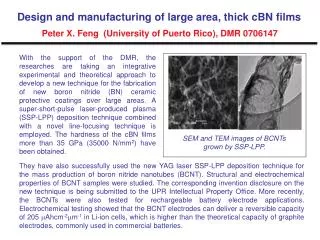
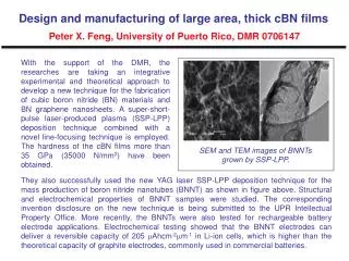
Download
1 / 33
340 likes | 391 Views
Polymer Analyses GPC, TEM, SEM. Speaker : 王睿詡 Date : 2009/11/23. Outline. GPC (Gel Permeation Chromatography) TEM (Transmission Electron Microscopy) SEM (Scattering Electron Microscopy). GPC (Gel Permeation Chromatography, Size Exclusion Chromatography). A Kind of LC Mechanisms.

E N D
Polymer AnalysesGPC,TEM, SEM Speaker : 王睿詡 Date : 2009/11/23
Outline • GPC(Gel Permeation Chromatography) • TEM (Transmission Electron Microscopy) • SEM (Scattering Electron Microscopy)
GPC (Gel Permeation Chromatography, Size Exclusion Chromatography)
A Kind of LC Mechanisms • Gel-Permeation Chromatography is a • mechanical sorting of molecules based • on the size of the molecules in solution. • Small molecules are able to permeate • more pores and are, therefore, retained • longer than large molecules. LC Separation Mechanisms Ref. : Rubinson and Rubinson, Contemporary Instrumental Analysis, Prentice Hall Publishing.
Filter (ca 0.5 um) GPC Columns Pump Injector Loop To Waste Concentration Detector Optional Detector #1 e.g. Viscosity Optional Detector #2 e.g Light Scattering To Waste To Waste To Waste Flow Voids: Uniform Diameter D helps minimize voids Pore: Size is given by the correlation length x x D = 1 - 50 mm Structural Picture
Column of GPC Hydrodynamic radius (Stoke’s radius) • K = cS /cM = (Ve - Vo)/Vi, since Vt = Vi + Vo + VG • (Vi is the volume of solvent held in its pores) • (Vg is the volume occupied by the solid matrix of the gel) • (Vo is the free volume outside the gel particles.) • K is distribution coefficient, we assume that • VG ≈ 0 • Both are normally values between 0 and 1 • Molecules that are too big to penetrate any of the pores elute in Vo and give K = 0 • Molecules that are small enough to penetrate all of the pores elute in Vi + Vo and give K = 1 • Between 0 and 1, larger molecules give smaller K values. • If a molecule gives a K larger than 1, it is likely interacting with the gel beads themselves (adsorption).
Common Detectors • UV – one of most common • Fluorescence – much greater sensitivity than UV • Refractive Index – widely used general detector • Electrochemical – based on amperometry, polarography, coulometry, • or conductometry. High sensitivity, wide applicability range • Mass Spectrometry – becoming increasingly used since interfacing • problems figured out. Expensive.
GPC Analysis Cheng-Jyun Huang and Feng-Chih Chang* Macromolecules 2008, 41, 7041-7052
GPC Analysis Chih-Feng Huanga, Hsin-Fang Leea, Shiao-Wei Kuoa, Hongyao Xua,b, Feng-Chih Changa,* Polymer 45 (2004) 2261–2269
GPC Analysis Chih-Feng Huang, Shiao-Wei Kuo, Hsin-Fang Lee, Feng-Chih Chang* Polymer 46 (2005) 1561–1565
TEM (Transmission Electron Microscopy) SEM (Scanning Electron Microscopy)
Size !! 肉眼可見物體的範圍:~10-3m 光學顯微鏡(OM)可見的範 圍:~10-6m 掃描式電子顯微鏡(SEM)可 見的範圍: ~10-8m (~10 nm) 穿透式電子顯微鏡(TEM)可 見的範圍: ~10-10m (~0.1 nm)
TEM (Transmission Electron Microscopy) From 交通大學貴儀中心
TEM (Transmission Electron Microscopy) • When E0-ΔEvalues become smaller, the Images become darker; When E0-ΔEvalues • become bigger, the Images become lighter. http://www.jeol.com/tem/jem2100f.html
Scanning Electron Microscope (SEM) From 交通大學貴儀中心
Structural Image of SEM http://cem.ess.nthu.edu.tw/K12/Article.asp?ColId=7806A63102F19
Principle of Imaging 0~50 ev
EDX(S) and WDX(S) http://en.wikipedia.org/wiki/Wavelength_dispersive_X-ray_spectroscopy
Images are Different Between in TEM and SEM SEM Image OM Image TEM Image
Material Science Angew. Chem. Int. Ed. 2002, 41, 688
Self-organization Morphology Solution state Bulkstate N : polymerization index χ: Flory-Huggins interaction parameter ƒ A : volume fraction of block A in the block copolymer Angew. Chem. Int. Ed. 2002, 41, 688
TEM (Transmission Electron Microscopy) Ref : Wan-Chun Chen,† Shiao-Wei Kuo,*,‡ U-Ser Jeng,§ and Feng-Chih Chang*,† Macromolecules 2008, 41, 1401-1410
TEM (Transmission Electron Microscopy) Ref : Shiao-Wei Kuo a,*, Pao-Hsaing Tung b, Feng-Chih Chang b European Polymer Journal 45 (2009)
TEM (Transmission Electron Microscopy) Ref : Chu-Hua Lu,† Chih-Feng Huang,† Shiao-Wei Kuo,‡ and Feng-Chih Chang*,† Macromolecules 2009, 42, 1067-1078
TEM (Transmission Electron Microscopy) Ref : Yuung-Ching Sheen a, Chu-Hua Lu a, Chih-Feng Huang a, Shiao-Wei Kuo b, Feng-Chih Chang a,* Polymer 49 (2008) 4017–4024
Scanning Electron Microscope (SEM) Y.-C. Yen et al. Polymer 49 (2008) 3625–3628 SHIH-CHI CHAN,1 SHIAO-WEI KUO,1 HWO-SHUENN SHE,2 HO-MAY LIN,1 HSIN-FANG LEE,1 FENG-CHIH CHANG1 Journal of Polymer Science: Part A: Polymer Chemistry, Vol. 45, 125–135 (2007)
Scanning Electron Microscope (SEM) Pao-Hsaing Tung, Shiao-Wei Kuo,*Shih-Chien Chan, Chih-Hao Hsu, Chih-Feng Wang, Feng-Chih Chang* Macromol. Chem. Phys. 2007, 208, 1823–1831
Scanning Electron Microscope (SEM) Chih-Feng Wang,1 Shih-Feng Chiou,2 Fu-Hsiang Ko,3 Cheng-Tung Chou,2 Han-Ching Lin,1 Chih-Feng Huang,1 Feng-Chih Chang*1; Macromol. Rapid Commun. 2006, 27, 333–337
Conclusion • Micro-Structural Analysis– TEM, SEM • Molecular Weight Determination - GPC