Download
1 / 34
380 likes | 817 Views
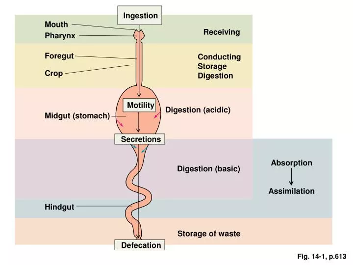
Ingestion. Mouth. Receiving. Pharynx. Foregut. Conducting Storage Digestion. Crop. Motility. Digestion (acidic). Midgut (stomach). Secretions. Absorption Assimilation. Digestion (basic). Hindgut. Storage of waste. Defecation. Fig. 14-1, p.613. Four basic digestive processes.

E N D
Ingestion Mouth Receiving Pharynx Foregut Conducting Storage Digestion Crop Motility Digestion (acidic) Midgut (stomach) Secretions Absorption Assimilation Digestion (basic) Hindgut Storage of waste Defecation Fig. 14-1, p.613
Four basic digestive processes • Motility: muscular contractions within gut tube that mix and move forward the contents of the digestive tract • Secretion: secretion of mucus, water, electrolytes, enzymes, • Digestion: broken down structurally complex foodstuffes into smaller, absorbable units • Absorption: transfer absorbable units from digestive tract lumen into the blood or hemolymph or body cavity.
+ + + + + Cerebral cortex Other inputs Salivary center in medulla Conditioned reflex Pressure receptors and chemoreceptors in mouth Autonomic nerves Simple reflex Salivary glands Salivary secretion Fig. 14-6, p.622
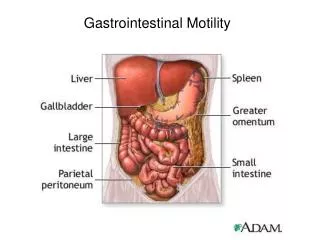
Esophagus Fundus Smooth muscle Gastroesophageal sphincter Body Stomach folds Pyloric sphincter Oxyntic mucosa Pyloric gland area Antrum Duodenum Fig. 14-8, p.627
Three main function of stomach • Store ingested food • Chemical digestion with HCl and enzymes • Produce chyme (a thick, liquid mixture)
Bile duct from liver Stomach Duodenum Hormones (insulin, glucagon) Blood Endocrine portion of pancreas (islets of Langerhans) Acinar cells secrete digestive enzymes Duct cells secrete aqueous NaHCO3 solution Exocrine portion of pancreas (acinar and duct cells) The glandular portions of the pancreas are grossly exaggerated. Fig. 14-12, p.638
Acid in duodenal lumen Fat and protein products in duodenal lumen Secretin release from duodenal mucosa CCK release from duodenal mucosa Neutralizes Digests + + Pancreatic duct cells Pancreatic acinar cells Secretion of aqueous NaHCO3 solution into duodenal lumen Secretion of pancreatic digestive enzymes into duodenal lumen (secretin carried by blood) (CCK carried by blood) Fig. 14-13, p.640
Negatively charged H2O-soluble portion (a carboxyl group at the end of a glycine or taurine chain) Small lipid (fat) droplet with bile salt molecules adsorbed on its surface Lipid-soluble portion (derived from cholesterol) Large fat droplet Through action of bile salts Lipid emulsion Fig. 14-17, p.644
Transverse colon Haustra Taeniae coli Descending colon Ascending colon Ileocecal valve Appendix Sigmoid colon Cecum Rectum External anal sphincter (skeletal muscle) Internal anal sphincter (smooth muscle) Anal canal Fig. 14-26, p.657
Rumen Reticulum Omasum Abomasum To small intestine (b) Fig. 14-27b, p.661