Download

1 / 126

E N D
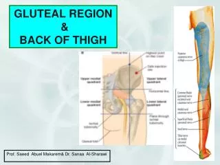
Place the cadaver in the prone position.Palpate again the following bony landmarks: the iliac crests, which end in front at the anterior superior iliac spine and behind at the posterior superior iliac spine. Identify the iliac tubercle, the ischial tuberosity, the greater trochanter of the femour, and the tip of the coccyx. Reexamine the landmarks on yourself.
Make a horizontal skin incision from the midline posteriorly along the upper border of the iliac crest to the iliac tubercle. If the perineum has not been dissected, make a vertical midline incision down the back of the lower lumbar region, sacrum, and coccyx, and encircle the anus.
Now make a transverse incision on the back of the thigh about 5 cm below the fold of the buttock. Connect the incision so that the skin flap and subcutaneous tissue can be turned laterally.
Cutaneous Nerves. Now remove the thick layer of superficial fascia along the lines of the skin incisions, starting in the area of the posterior superior iliac spine and working downward and laterally.
When removing the fascia, attempt to identify the cutaneous branches of the upper three lumbar and upper three sacral nerves as they cross the posterior part of the iliac crest. Also identify the lateral branches of the iliohypogastric and twelfth thoracic nerves as they cross the anterior part of the iliac crest.
Preserve the branches from the posterior cutaneous nerve of the thigh (posterior femoral cutaneous nerve ) and, if possible, those from the lateral cutaneous nerve of the thigh ( lateral femoral cutaneous nerve).
Gluteal Muscles.Clean the deep fascia covering the gluteus maximus and gluteus medius muscles. Carefully define the upper and lower borders of the gluteus maximus muscle and, without damaging the underlying structures, incise the deep fascia along these borders.
Now insert your fingers beneath the upper and lower borders of this muscle and separate it from the deep structures. With a curved incision, cut the gluteus maximus free from its origin, starting in the region of the posterior superior iliac spine. Reflect the muscle laterally to its insertion into the iliotibial tract and the femur.
Avoid damaging the underlying gluteus medius muscle and the sacrotuberous ligament. Identify and clean the inferior gluteal nerve and vessels as they enter the deep surface of the gluteus maximus. Identify the bursae over the greater trochanter and the ischial tubersity.
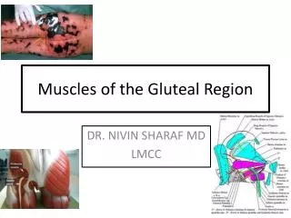
Identify and clean the following muscles in the gluteal region from above downward:1. The tensor fasciae latae as it passes downward from the anterior superior iliac sipne to be inserted into the iliotibial tract.
2. The gluteus medius muscle, whose fibers converge to be inserted intothe lateral surface of the greater trochanter.3. The piriformis muscle, which emerges from the greater sciatic foramen and is inserted into the upper border of the greater trochanter.
Cut through the origin of the gluteus medius and reflect it downward and laterally to its insertion on the greater trochanter.
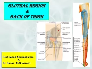
Nerves and Blood Vessels of the Gluteal Region.Identify, clean, and study the following nerves and vessels in the gluteal region, using the piriformis muscle as the central key structure.
At the superior border of the piriformis, identify:1. The superior gluteal nerve, which emerges from the greater sciatic foramen and supplies the gluteus medius and minimus and the tensor fasciae latae muscles.
2. The superior gluteal artery, a branch of the internal iliac artery that emerges from the greater sciatic foramen to enter the gluteal region.
At the lower border of the piriformis muscle, identify:1. The sciatic nerve, the largest nerve in the body, which emerges from the greater foramen to pass downward into the back of the thigh.
Occasionally, the peroneal part of the sciatic nerve leaves the sciatic nerve high in the pelvis and appears in the gluteal region by passing above or through the piriformis muscle. The sciatic nerve ususlly gives no branches in the gluteal region.
2. The inferior gluteal nerve, which also emerges from the greater sciatic foramen and supplies the gluteus maximus muscle.3. The inferior gluteal artery, a branch of the internal iliac artery, which also emerges from the greater sciatic foramen.
4. The posterior cutaneous nerve of the thigh, which passes downward from the greater sciatic foramen on the posterior surface of the sciatic nerve. It leaves the gluteal region by passing superficial to the biceps femoris.
Identify the gluteal branches to the skin over the lower medial quadrant of the buttock and the perineal branch, which passes medial to the scrotum or labium majus.
5. The pudenal nerve, the nerve to the obturator internus, and the internal pudendal artery, which enter the gluteal region briefly through the greater sciatic foramen. They cross the ischial spine and then enter the perineum through the lesser sciatic foramen. The nerve to the obturator internus gives off a small branch that supplies the superior gemellus muscle.
6. The nerve to the quadratus femoris, which leaves the pelvis through the greater sciatic foramen and runs downward anterior to the sciatic nerve, the gemelli,and the tendon of the obturator internus. Identify its branches to the inferior gemellus and the quadratus femoris muscles.
Where necessary, remove the veins that accompany the arteries in order to clarify your dissection.
It is most important that you confirm the position of the sciatic nerve in relation to the greater trochanter and the ischial tuberosity. Replace the gluteus medius and maximus muscles in their correct anatomical position.
Now relate the sciatic nerve to the gluteus maximus muscle; remember that a large number of intramuscular injections are introduced into this muscle and that the sciatic nerve must not be damaged. Note that the upper lateral quadrant of this muscle lies a safe distance away from the sciatic nerve.
Clean the ischial tuberosity and note the attachment of the sacrotuberous ligament and the origins of the hamstring muscles.
Make a transvers skin incision at the level of the midcalf. Make a vertical skin incision down the midline on the back of the thigh, connecting the transverse incision below the buttock to the transverse incision on the midcalf.
Reflect the skin flaps medially and laterally. Try to identify some of the branches of the posterior cutaneous nerve of the thigh along the middle of the back of the thigh. Remove the superficial fascia.
Incise in the midline the deep fascia on the back of the thigh from the gluteal region to the calf. Reflect the cut edges medially and laterally.
Identify and clean the hamstring muscles, namely, the biceps femoris, the semimembranosus, and the semitendinosus. Confirm that these muscles arise from the ischial tuberosity. Identify the short head of the biceps, which arises from the shaft of the femur.
Sciatic Nerve. Follow the sciatic nerve from the gluteal region deep to the long head of the biceps femoris. Note that it descends in the midline of the thigh, that it is overlapped posteriorly by the adjacent margins of the biceps femoris and semimembranosus muscles, and that it lies on the posterior aspect of the adductor magnus muscle.
In the lower third of the thigh the sciatic nerve divides into the tibial and common peroneal nerve. Occasionally the sciatic nerve divides into its two terminal branches at a higher level.
Identify the following branches of the sciatic nerve:1. Muscular branches to the long head of the biceps femoris, the semitendinosus, the semimembranosus, and the ischial fibers (hamstring part) of the adductor magnus. These branches arise from the tibial component of the sciatic nerve and run medially, a short distance below the ischial tuberosity, to supply the muscles.
2. Tibial Nerve.3. Common peroneal nerve.Detach the hamstring muscles from the ischial tuberosity and fully expose the posterior surface of the adductor magnus. Define the insertion of the muscle into the femur. Identify the four perforating arteries, which are branches of the profunda femoris that pierce the adductor magnus.
Trace the tendons of the sartorius, gracilis, and semitendinosus muscles to their insertions on the medial surface of the shaft of the tibia.
Identify and clean the boundaries of the popliteal fossa. Note that the upper lateral pboundary is formed by the biceps femoris and that the upper medial boundary is formed by the semitendinosus and semimembranosus muscles. The lower boundaries are formed by the medial and lateral heads of the gastrocnemius muscle.
Separate the medial and lateral heads of the gastrocnemius muscle and identify the plantaris muscle. Expose and clean the common peroneal and tibial nerves and their branches.
Common Peroneal Nerve. Having arisen from the sciatic nerve, the common peroneal nerve runs downward through the popliteal fossa, following the medial border of the biceps femoris muscle. Note that it leaves the fossa by crossing superficially the lateral head of the gastrocnemius muscle. It then passes posterior to the head of the fibula and winds laterally around the neck of the bone to enter the substance of the peroneus longus muscle.
Identify the following branches of the common peroneal nerve:1. Cutaneous a. The peroneal communicating nerve, which runs downward and joins the medial sural cutaneous nerve to form the sural nerve.b. The lateral sural cutaneous nerve, which supplies the skin on the lateral side of the back of the leg.
2. Muscular branches to the short head of the biceps femoris muscle.3. Articular branches to the knee joint.
Tibial Nerve. Follow this nerve from its origin from the sciatic nerve, downward through the popliteal fossa. It lies first on the lateral side of the popliteal artery, then posterior to it, and finally medial to it. Note that the popliteal vein lies between the nerve and the artery through its course. The tibial nerve leaves the fossa by passing beneath the soleus muscle.
Identify the following branches of the tibial nerve:1. Cutaneous. The medial sural cutaneous nerve descends between the two heads of the gastrocnemius muscle and is usually joined by the peroneal communicating branch of the common peroneal nerve to form the sural nerve.
2. Muscular branches. These supply both heads of the gastrocnemius and the plantaris, soleus, and popliteus. Note that the nerve to the popliteus runs down over the muscle and then winds around the lower border to supply the popliteus on its anterior surface.3. Articular branches to the knee joint.
Now Clean the popliteal artery and vein.Popliteal Artery. Trace the popliteal artery through the popliteal fossa. Note that it begins as a continuation of the femoral artery at the opening in the adductor magnus. The artery ends at the lower border of the popliteus by dividing into anterior and posterior tibial arteries.