Download

1 / 34
370 likes | 530 Views
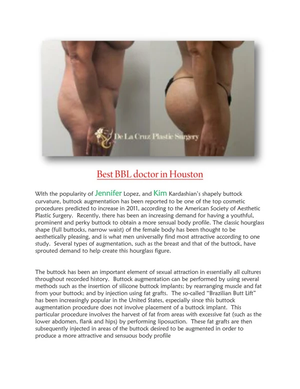
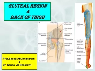
The Gluteal Region (Buttock). Dr. Zeenat Zaidi. Gluteal Region. It is the region behind the pelvis, extending from the iliac crest superiorly to the gluteal fold (fold of the buttock) inferiorly

E N D
The Gluteal Region (Buttock) Dr. Zeenat Zaidi
Gluteal Region • It is the region behind the pelvis, extending from the iliac crest superiorly to the gluteal fold (fold of the buttock) inferiorly • Gluteal fold indicates the lower border of the gluteus maximus muscle (glutealsulcus/crease is a skin crease for the hip joint) • A deep midline groove, the natal (intergluteal) cleft separates the buttocks from each other. Natal cleft Buttock Gluteal crease Gluteal fold
Superficial fascia • Thick, dense, well developed, laden with large quantities of fat (specially in women) that: • Gives the characteristic convexity to the buttock • Forms a thick cushion over the ischialtuberosity Natal cleft Buttock Gluteal crease
Dermatomes Cutaneous Nerve supply: • Upper lateral quadrant: Lateral branches of iliohypogastric (L1) and T12 • Upper medial quadrant: Posterior rami of L1,2,3 & S1,2,3 • Lower lateral quadrant: branches from lateral cutaneous nerve of thigh (L2,3) • Lower medial quadrant: branches from posterior cutaneous nerve of thigh (S1,2,3) • Skin in the floor of the natal cleft: branches from lower sacral and coccygeal nerves
Superficial Inguinal lymph nodes • The skin and the fat of the gluteal region is: • Supplied by perforating branches of the superior and inferior gluteal arteries • Drain into the lateral group of the superficial Inguinal lymph nodes
Deep Fascia Fascia over gluteus medius • Is continuation of the fascia lata (deep fascia of the thigh) • At the lower border of the gluteus maximus, fascia lata splits to enclose the muscle • Above the gluteus maximus, the deep fascia continues as one layer covering the gluteus medius& gets attached to iliac crest • Laterally the fascia merges with the iliotibial tract Tensor fascia lata Gluteal fascia Iliotibial tract
The gluteal region contains: • Bones • Ligaments • Muscles • Vessels • Nerves
Bones of the Gluteal Region • Posterior aspect of: • Hip bone • Femur & • Hip joint
Ligaments of the Gluteal Region • 2 ligaments: • Sacrospinous, connecting sacrum to ischial spine • Sacrotuberous, connecting sacrum to ischialtuberosity • They convert the greater & lesser sciatic notches into greater & lesser sciatic foramina • Their main function is to: • Stabilize the sacrum • Prevent its posterior rotation at the sacroiliac joint
Structures passing through the greater sciatic foramen Above the piriformis: Superior gluteal vessels & nerve Piriformis: an important landmark Below the piriformis: Inferior gluteal vessels & nerve Sciatic nerve Posterior cutaneous nerve of thigh Pudendalnerve & Internal pudendal vessels Nerve to obturator internus Nerve to quadratus femoris
Structures passing through the lesser sciatic foramen Entering: Pudendal nerve & Internal pudendal vessels Exiting: Tendon of obturator internus Nerve to obturator internus
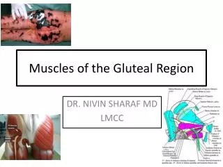
Muscles of the Gluteal Region • Gluteus maximus • Gluteus medius • Gluteus minimus • Tensor fascia lata • Piriformis • Superior Gemellus • Inferior Gemellus • Obturatorinternus • Quadratusfemoris
Gluteus Maximus • Largest muscle in the body • Forms the prominence of buttock • Origin: • Outer surface of ilium behind the posterior gluteal line • Lumbar fascia • Posterior surface of sacrum & coccyx • Sacrotuberous ligament ilium S C
Gluteus maximus Iliotibial tract • Insertion: • Most of the muscle (3/4th) inserted into the iliotibial tract • Deeper fibers inserted to the glutealtuberosity • Nerve supply: • Inferior gluteal nerve (L5, S1, 2)
Actions: • Extends & laterally rotates the hip joint • Extends the knee joint (through iliotibial tract) • Gives simultaneous stability to the hip and knee joints through the iliotibial tract Gluteus maximus is the chief antigravity muscle of the hip. It is used in standing up from a sitting position, running & climbing up stairs. In each case extension of the hip moves the trunk upwards. The muscle must be extremely powerful to raise the weight of the body against gravity. This is called "forced extension".
Gluteus Medius • Origin: outer surface of ilium between the middle and posterior gluteal lines • Insertion: Lateral surface of greater trochanter • Nerve supply: Superior gluteal nerve (L4,5, S1) • Action: • Abducts & medially rotates the thigh • Steady pelvis in walking
Gluteus Minimus • Origin: outer surface of ilium • Insertion: Anterior surface of greater trochanter • Nerve supply: Superior gluteal nerve (L4,5, S1) • Action: Abducts & medially rotates the thigh
Tensor Fascia Lata Tensor fascia lata • Origin: Outer edge of iliac crest between anterior superior iliac spine & iliac tubercle • Insertion: Into the iliotibial tract • Nerve supply: Superior gluteal nerve (L4,5, S1) • Action: Maintains the knee in extended position Iliotibial tract
Piriformis • Origin: Anterior surface of S2,3,4 vertebrae • Insertion: Upper border of greater trochanter • Nerve supply: Anterior rami of S1,2 • Action: • Lateral rotator of thigh • Assists in stabilizing hip joint especially in abduction Piriformis forms an important landmark in the region
Obturator Internus • Origin: Inner surface of obturator membrane and adjacent bone • Insertion: Upper border of greater trochanter along with gemelli • Nerve supply: nerve to obturatorinternus (L4,S1) • Action: Lateral rotator of thigh
Superior & Inferior Gemelli • Origin: • Superior from ischial spine • Inferior from ischialtuberosity • Insertion: Upper border of greater trochanter • Nerve supply: • Superior from nerve to obturatorinternus (L4, S1) • Inferior from nerve to quadratusfemoris (L4, S1) • Action:Lateral rotators of thigh
Quadratus Femoris • Origin: Lateral border of ischialtuberosity • Insertion: Quadrate tubercle of femur • Nerve supply: nerve to quadratusfemoris (L4,S1) • Action: Lateral rotator of thigh
Nerves of the Gluteal Region • Sciatic • Posterior cutaneous nerve of the thigh • Superior gluteal • Inferior gluteal • Nerve to quadratusfemoris • Pudendal nerve • Nerve to obturatorinternus
Arteries of the Gluteal Region • Branches of internal iliac artery: • Superior gluteal • Inferior gluteal • Branches of femoral artery: • Lateral circumflex • Medial circumflex • Branche of profundafemoris artery: • First perforating branch
Superior & Inferior Gluteal Arteries • Are branches of the internal iliac artery • Enter the gluteal region through the greater sciatic foramen (superior gluteal artery above the piriformis, inferior gluteal artery below the piriformis) • Supply the gluteal region and contribute to the anastomosis around the hip joint
Trochanteric Anastomosis • Is the main supply to the head & neck of femur • Provides a connection between internal iliac and femoral arteries • Lies near the trochantericfossa, branches run along the femoral neck beneath the reticular fibers of the capsule • Formed by: • Descending branches of superior and inferior gluteal arteries & • Ascending branches of lateral and medial circumflex arteries
Arterial supply to Femoral head • Medial & lateral femoral circumflex arteries • Superior and inferior gluteal arteries • Post. obdurator artery via artery of femoral ligament TROCHANTERIC ANASTOMOSIS Posterior view
Cruciate Anastomosis • Lies at the level of lesser trochanter • Provides a connection between internal iliac and femoral arteries • Formed by: • Descending branch of inferior gluteal artery • Transverse branches of medial and lateral circumflex arteries & • Ascending branch of first perforating artery
Bursae Related to Gluteus Maximus • Gluteofemoral Bursa: lies between gluteus maximus tendon and vastuslateralis • Trochanteric Bursa: liesbetween gluteus maximus tendon and greater trochanter • Ischial Bursa: liesbetween gluteus maximus & ischialtuberosity
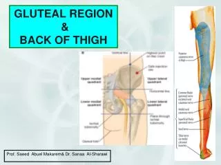
Safe Area for Intramuscular Injection • Intramuscular injection enables a large amount of a drug to be introduced at once but absorbed gradually. • The injection site must be carefully selected to avoid injury to the underlying large vessels and nerves. Outer upper quadrant of the buttock is the safe area for intramuscular injection to avoid injury to the underlying sciatic nerve
Trendelenburg Test • Observe patient from behind, ask him/her to stand on one foot and then the other • Negative test: Pelvis ‘tilts up’ on contralateral side • Positive test: Pelvis ‘sags’ on contralateral side • To assesses whether the hip abductors (particularly gluteus medius) are functioning normally
Problems that could lead to a positive Trendelenburg test: • Fracture neck of femur • Dislocation of hip joint • CoxaVara • Nonfunctioning gluteus medius and minimus due to: • Neurological damage (L4 – 5 disc herniation) • Any disease affecting muscles (myopathy)