Download
1 / 13
130 likes | 285 Views
The Movement of Blood. There are two distinct circuits of blood flow in the human body. Heart. Double Pump Septum separates left and right sides of heart Right side receives blood from body and pumps to the lungs (deoxygenated)

E N D
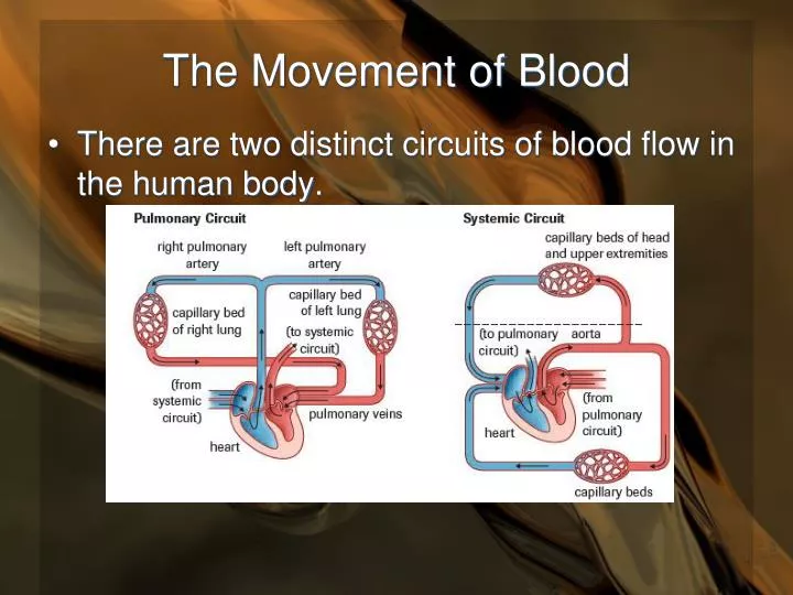
The Movement of Blood • There are two distinct circuits of blood flow in the human body.
Heart • Double Pump • Septum • separates left and right sides of heart • Right side receives blood from body and pumps to the lungs (deoxygenated) • Left side receives blood from lungs and pumps to the body (oxygenated) • Left side has thicker walls than right side • must pump with greater force - longer distance
Heart Atria • Receive blood from veins • Pump blood to ventricles • Walls are relatively thin • Vena cava (R); Pulmonary vein (L) • No valves between veins and atria
Heart • Ventricles • Receive blood from atria • Pump blood into arteries • Left: aorta; Right: pulmonary artery • Thick, muscular walls (especially left side) • pump blood throughout body (left) • Semilunar valves between ventricles and arteries
Coronary arteries • These arteries supply the heart with O2. • The heart can use up to 20% of blood O2 during stress. It can never stop working • Coronary bypass surgery adds a blood vessel which bypasses a blocked coronary artery.
Heart • AV (atrioventricular) valves between atria and ventricles • Chordae tendonae and papillary muscles • Left: Bicuspid (Mitral) valve • Right: Tricuspid valve
1 23 4567 8 9 Deoxygenated blood from the BODY entering the heart The RIGHT ATRIUM The VALVES prevent blood flowing the wrong way when the atria and ventricles contract The RIGHT VENTRICLE The thicker walled LEFT VENTRICLE The LEFT ATRIUM Oxygenated blood FROM THE LUNGS entering the heart Oxygenated blood leaving the heart and flowing TO THE BODY Deoxygenated blood leaving the heart and flowing TO THE LUNGS
Control of Heartbeat • Myogenic Muscle • “self-stimulating” • Pacemaker - SA (sino-atrial) node • located in right atrium • Initiates heartbeat and sets rate • Causes atria to contract
Control of Heartbeat • AV (Atrioventricular) node • Located in right atrium near ventricle • Receives impulse from SA node • Conducts impulse through septum to apex of heart via special nervous tissue (Purkinje fibers)
Control of Heartbeat • Nerve fibers radiate from apex of heart up through the ventricles • Ventricles contract as impulse moves through these fibers • Contraction from the apex up pushes blood up to arteries
Control of Heartbeat • Heart rate is influenced by nervous system Autonomic nerves 1. Sympathetic nerves • Speed up heart rate 2. Parasympathetic nerves • Slow down heart rate • Tachycardia (fast); Bradycardia (slow)
Heart Sounds • “Lubb Dubb” • Lub (first sound) caused by closing of AV valves • Dub (second sound) caused by closing of semilunar valves