Download
1 / 39
420 likes | 732 Views
DEPARTMENT OF ANATOMY. Lower limb. WINDSOR UNIVERSITY SCHOOL OF MEDICINE . Dr. SREEKANTH THOTA. Overview of the Lower Limb. Regions of the lower limb.

E N D
DEPARTMENT OF ANATOMY Lower limb WINDSOR UNIVERSITYSCHOOL OF MEDICINE Dr. SREEKANTH THOTA
Regions of the lower limb. • The lower limb is divided into the gluteal region, thigh, leg, and foot on the basis of major joints, component bones, and superficial landmarks.
Areas of transition • The femoral triangle and popliteal fossa, and the posteromedial side of the ankle are important areas of transition through which structures pass between regions .
Lower limb Schedule • Lecture • 1:Skin,Superficial fascia, deep fascia, cutaneous veins and nerves • 2:Femoral triangle and adductor canal • 3:Ant compt of thigh • 4: Medial compt of thigh • 5:Gluteal region • 6: Post compt of thigh • 7: Popliteal fossa • 8: Ant compt of leg • 9: Post compt of leg
cont • 10. Lateral compt of leg • 11.Dorsum of foot • 12.Sole • 13.Review of lower limb
Fascia of the Lower Limb • Superficial fascia : • consists of loose connective tissue that contains a variable amount of fat, cutaneous nerves, superficial veins (great and small saphenous veins and their tributaries), lymphatic vessels, and lymph nodes.
Deep fascia • Fascia lata The outer layer of deep fascia in the lower limb forms a thick 'stocking-like' membrane, which covers the limb and lies beneath the superficial fascia . This deep fascia is particularly thick in the thigh and gluteal region and is termed the fascia lata. Inferiorly, the fascia lata is continuous with the deep fascia of the leg
Iliotibial tract • The fascia lata is thickened laterally into a longitudinal band (the iliotibial tract), which descends along the lateral margin of the limb from the tubercle of the crest of the ilium to a bony attachment just below the knee. • This broad band of fibers is the conjoint aponeurosis of the tensor of fascia lata and gluteus maximus muscles.
Superficial Veins of the Lower Limb • Superficial Veins • The two major superficial veins in the lower limb are the great and small saphenous veins. • Are in subcutaneous tissue • Normally send blood to deep veins via perforating veins • Deep veins • Are deep to deep fascia and accompany all major arteries • Superficial and deep veins have valves, which are more numerous in deep veins.
perforating veins • Contain valves that allow blood to flow only from the superficial veins to the deep veins.
great saphenous vein • Formed by the union of the dorsal vein of the great toe and the dorsal venous arch of the foot. • Ascends anterior to the medial malleolus. • Passes posterior to the medial condyle of the femur
great saphenous vein • Traverses the saphenous opening in the fascia lata. • Empties into the femoral vein.
Small saphenous vein • Arises on the lateral side of the foot from the union of the dorsal vein of the little toe with the dorsal venous arch. • Ascends posterior to the lateral malleolus • Ascends between the heads of the gastrocnemius muscle. • Empties into the popliteal vein in the poplitealfossa.
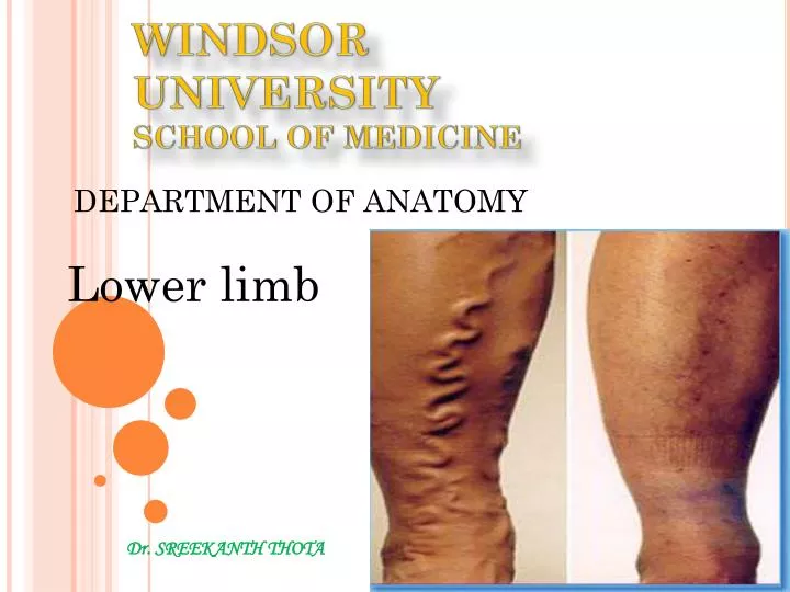
Varicose Veins • Varicose veins are veins that have become enlarged and tortuous. • Varicose veins form when the valves that usually prevent blood flow from the deep veins through the perforating veins to the superficial veins are incompetent.
Varicose Veins • Varicose veins are common in the posteromedial parts of the lower limb. • Causes • More common in women than in men, and are linked with heredity. • Prolonged standing • pregnancy, obesity, menopause, aging etc
Deep vein thrombosis (DVT) • Formation of a blood clot, known as a thrombus, in the deep leg vein. • Deep vein thrombosis can develop if you're sitting still for a long time, such as when traveling by airplane or car. • Serious condition because a blood clot that has formed in your vein can break loose and travel to your lungs(life-threatingpulomnary embolism).
Pulmonary Embolism • Left untreated, a deep vein thrombosis (DVT) can break off and travel in the circulation, getting trapped in the lung, where it blocks the oxygen supply, causing heart failure. Deep Vein Thrombosis {DVT} >>> IVC >>> RA>>> RV>>> Pulmonary Trunk>>> Branch of Pulmonary Artery blocked >>> Death of Lung tissue
Saphenous Vein Grafts • Great saphenous vein is commonly used for coronary arterial bypasses because • 1. Readily accessible • 2. Sufficient distance occurs between the tributaries and the perforating veins so that usable lengths can be harvested • 3. Wall contains a higher percentage of muscular and elastic fibers than do other superficial veins.
Saphenous Vein Grafts • When part of the great saphenous vein is removed for a bypass, the vein is reversed so that the valves do not obstruct blood flow in the graft.
Lumbar Plexus • The lumbar plexus of nerves is formed anterior to the lumbar transverse processes, within the proximal attachment of the psoas major. • This nerve network is composed of the anterior rami of L1 through L4 nerves.
The following nerves are branches of the lumbar plexus • 1. Iliohypogastric nerve(L1) • 2. Ilioinguinal nerve(L1) • 3. Lateral cutaneous nerve of the thigh: (L2, L3)Skin of anterior and lateral surfaces of the thigh • 4. Genitofemoral nerve (L1, 2): Cremaster muscle in scrotum in male; skin over anterior surface of thigh; nervous pathway for cremasteric reflex.
Ilioinguinal nerve(L1) In the male ("anterior scrotal nerve"): to the skin over the root of the penis and upper part of the scrotum. In the female ("anterior labial nerve"): to the skin covering the mons pubis and labium majus.
Lateral cutaneous nerve of thigh Cutaneous nerve that innervates the skin on the lateral part of the thigh.
Genitofemoral nerve (L1, 2) The genitofemoral nerve is responsible for both the sensory (femoral branch) and motor portions (genital branch) of the cremasteric reflex, which describes contraction of the cremasteric muscle when the skin of the superior medial part of the thigh is touched.
Cremasteric reflex • The cremasteric reflex is a reflex in human males. • The cremasteric reflex is dependent upon the nerve roots L1 and L2 • This reflex is elicited by lightly stroking the superior and medial part of the thigh in a downward direction. The normal response is a contraction of the cremaster muscle that pulls up the scrotum and testis on the side stroked. • Upper and lower motor neurone disorders can cause an absence of the cremasteric reflex.
5. Femoral nerve (L2, 3, 4): Iliacus, pectineus, sartorius, quadriceps femoris muscles, and intermediate cutaneous branches to the skin of the anterior surface of the thigh and by saphenous branch to the skin of the medial side of the leg and foot; articular branches to hip and knee joints
6. Obturator nerve (L2, 3, 4): Gracilis, adductor brevis, adductor longus, obturator externus, pectineus, adductor magnus (adductor portion), and skin on medial surface of thigh; articular branches to hip and knee joints. • The lumbosacral trunk (L4, L5) passes over the ala (wing) of the sacrum and descends into the pelvis to participate in the formation of the sacral plexus with the anterior rami of S1 to S4 nerves
Lymphatic Drainage of the Lower Limb • The lower limb has superficial and deep lymphatic vessels. • Superficial lymphatic vessels: • Superficial inguinal lymph nodes • Deep lymphatic vessels • Deep inguinal lymph nodes Accompany deep veins and enter the popliteal lymph nodes. • Popliteal nodes>>deep inguinal>external iliac>>internal iliac>>>common iliac