Download
1 / 28
280 likes | 433 Views
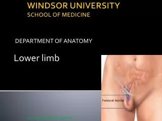
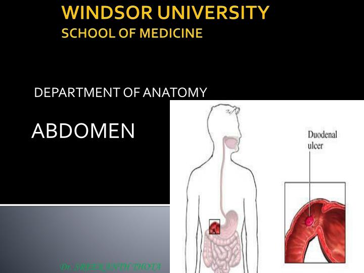
Dr. SREEKANTH THOTA. DEPARTMENT OF ANATOMY. ABDOMEN. WINDSOR UNIVERSITY SCHOOL OF MEDICINE. Small Intestine. The small intestine is the longest part of the gastrointestinal tract and extends from the pyloric orifice of the stomach to the ileocecal fold.

E N D
Dr. SREEKANTH THOTA DEPARTMENT OF ANATOMY ABDOMEN WINDSOR UNIVERSITYSCHOOL OF MEDICINE
Small Intestine The small intestine is the longest part of the gastrointestinal tract and extends from the pyloric orifice of the stomach to the ileocecal fold. This hollow tube, which is approximately 6-7 m long with a narrowing diameter from beginning to end, consists of the duodenum, the jejunum, and the ileum.
Duodenum • The duodenum curves in a C shape around the head of the pancreas and is 10 in (25 cm) long. • At its origin from the pylorus it is completely covered with peritoneum for about 1 in (2.5 cm), but then becomes a retroperitoneal organ.
Blood supply of the Duodenum • Superior & inferior pancreaticoduodenal arteries.
Jejunum and IleumTogether, the jejunum and ileum are 6 to 7 m long Although no clear line of demarcation between the jejunum and ileum exists, they have distinctive characteristics that are surgically important
Jejunum • The second part of the small intestine, the jejunum, begins at the duodenojejunal flexure where the alimentary tract resumes an intraperitoneal course. • The jejunum constituting approximately two fifths of the intraperitoneal section of the small intestine • Most of the jejunum lies in the left upper quadrant of the infracolic compartment
Ileum • The ileum, ends at the ileocecal junction, the union of the terminal ileum and the cecum • The ileum constituting approximately three fifths of the intraperitoneal section of the small intestine • ileum lies in the right lower quadrant
Distinguishing Characteristics of the Jejunum and Ileum in the Living Body
Superior mesenteric artery • Comes off from aorta just inferior to celiac trunk. • Origin is hidden behind the pancreas • Comes off between head of pancreas and uncinate process of pancreas and passes over 3rd part of duodenum to enter the root of mesentery.
Superior mesenteric artery branches • Branches • Inferior Pancreaticoduodenal. • Jejunal and ileal arteries • Ileocolic • Right Colic • Middle Colic
Braches of ileocolic: • Anterior cecal • Posterior cecal • Important branch of posterior cecal: Appendicular
Superior mesenteric vein • It lies anterior and to the right of the SMA in the root of the mesentery • The SMV ends posterior to the neck of the pancreas, where it unites with the splenic vein to form the portal vein
Innervation of small intestine Superior mesenteric nerve plexus The parasympathetic fibers in the nerves to the jejunum and ileum derive from the posterior vagal trunks. The sympathetic fibers in the nerves to the jejunum and ileum originate in the T10 to T11 segments of the spinal cord and reach the superior mesenteric nerve plexus through the sympathetic trunks