Download
1 / 22
350 likes | 1.18k Views
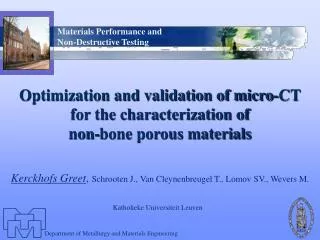
Characterization of porous scaffold materials for bone tissue engineering - Saartje Impens - . Micro-CT symposium 31/05/07. GBE project. 2 different aims: Setting up a protocol for the healing of large and complex, but critical bone defects

E N D
Characterization of porous scaffold materials for bone tissue engineering - Saartje Impens - Micro-CT symposium 31/05/07
GBE project • 2 different aims: • Setting up a protocol for the healing of large and complex, but critical bone defects • High throughput screening of different scaffolds (= porous structure) With the aid of micro-CT evaluation
1. Healing of critical bone defects Scaffold seeding and culturing with cells Scaffold Cells + medium Bioreactor Patient own cells + growth factors Operation Room Haeled bone defect Bone defect In vitro
2. High throughput screening input = material + coating + growth factors Optimize scaffold Toxicity testing Yes Clinical approved scaffold Not Ok No REJECT Ok If 2D plates are possible Optimization possible? Yes 2D plates 3D scaffold No µ-CT screening Macrostructural & Mechanical parameters Fluid Flow Macrostructural shortcoming Further screening until clinical approvement Not Ok Yes input = cells Ok No No perfusion perfusion possible 2D cell seeding 3D cell seeding in vivo screening nude mice 2D cell culture 3D cell culture No output = proliferation differentiation Yes time point analysis No Yes
GBE strategy • Multidisciplinary approach
Micro-CT use • Micro-CT based characterization of scaffolds • Calculate structural parameters • Calculate mechanical parameters with the aid of a FE-model • Calculate fluid flow • Evaluation of bone formation in explanted scaffold • Replacement of histology?
1. Scaffold characterization • Important parameters for bone formation in Matlab • Porosity As high as possible (100%) • Specific surface area As high as possible (Mentat) >3,95mm-1 (Ding et al. based on bone) • Pore size 100-800µm (PorousAnalyser) • Permeability As high as possible (PoreNet) > 10-8m2 (Kohles et al. based on bone) • Interconnectivity As high as possible (100%) • Mechanical parameters with FE-modeling (Mesh creation in Matlab) Expected load during walking is 1,2 x body weight • Strength 100% under yield strength • Stiffness 17-20 GPa (cortical bone) 10-1500MPa (trabecular bone) • Stretch on surface (500-)1500-4000µstrain
1. Scaffold characterization Scaffolds Reconstructed micro-CT FE-mesh Image
1. Scaffold characterization • Structural and biomechanical parameters
Scaffold characterization • Extra important parameter for the GBE project • Fluid flow • Nutrient & Oxygen transport • Wall shear stress • May stimulate proliferation and differentiation i.e. May stimulate bone formation • Ideally Computing Fluid Flow of micro- CT based models
1. Scaffold characterization • 2D Fluid flow on µCT based model Inflow: 1 ml/min Scaffold: Ø 6 mm, L 8 mm Figures: Tim van Cleynenbreugel
1. Scaffold characterization • 3D Fluid flow on CAD-based model Figures: Silvia Truscello
1. Scaffold characterization • Problems occur when meshing regular scaffolds produced by rapid prototyping Blue Best Violet Pink Orange Red Worst Manually remeshing
* 2. Substitute for Histology • Evaluation different scaffold materials • Time consuming • Embedding 2 weeks • Sectioning • 1 scaffold/day • Labor intensive • Staining • 1 day • Analysis • 1 scaffold/day • Labor intensive
2. Substitute for Histology • Polymer scaffolds Binarized histological Section Interpolated micro-CT image After registration Green: Overlap Blue: only histology Red: only micro-CT Histological image
2. Substitute for Histology • Distinguish between scaffold and bone by thresholding? Difficult, depends on scaffold material Bone Scaffold Zone of bone ingrowth
2. Substitute for Histology • Micro-CT analysis • Micro-CT Scanning • Micro-CT scanning explant • Positioning and subtrac- ting in Mimics to determine the amount of bone ingrowth
Conclusion • Micro-CT is a very useful tool for this type of research • Scaffold parameters can be calculated • Prior to implantation • Non destructive • Time consuming histology • Can be replaced • If necessary, histology can be performed after scanning • If FE models and meshing problems are solved • Fluid flow • Wall shear stresses can be calculated
Acknowledgement Special thanks goes to: • Jan Schrooten • Tim van Cleynenbreugel • Barbara Neirinck • Silvia Truscello • Greet Kerckhofs -Thanks-