Download
1 / 70
700 likes | 741 Views
ACUTE INFLAMMATION. H.A.MWAKYOMA, MD. History. • Egyptian papyrus - 3000 B.C. • Celsus (Roman in 1st century A.D.) Rubor - Tumor - Calor - Dolor redness - swelling - heat - pain • Virchow added functio laesa later. What is inflammation?. Inflammation –

E N D
ACUTE INFLAMMATION H.A.MWAKYOMA, MD
History • Egyptian papyrus - 3000 B.C. • Celsus (Roman in 1st century A.D.) Rubor - Tumor - Calor - Dolor redness - swelling - heat - pain • Virchow added functio laesa later
What is inflammation? • Inflammation – Protective response intended to eliminate the initial cause of cell injury and the necrotic cells and tissues arising from the injury • Inflammation is intimately associated with the repair process which includes parenchymal cell regeneration and scarring
Inflammation • Acute - minutes to days • Characterized by fluid and protein • PMN’s • Exudate SG > 1.020 • Chronic - weeks to years • Lymphocytes and macrophages • ACUTE Inf - PMN’s (Polymorphonuclear Cells) • CHRONIC Inf - Mononuclear Cells EXUDATE
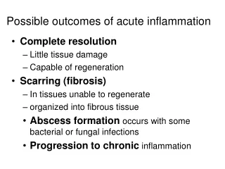
Acute inflammation “The immediate and early response to an injurious agent” Chronic inflammation “Inflammation of prolonged duration (weeks or months) in which active inflammation, tissue destruction, and attempts at repair are proceeding simultaneously“
Exudate • vascular permeability • high protein & cell debris • SG > 1.020 Transudate • normal vascular permeability • hydrostatic pres. plasma ultrafiltrate • low protein (mostly albumin) • SG < 1.012 Edema • exudate or transudate ; interstitium or cavity
Acute inflammation major components • Transient vasoconstriction • Vasodilatation • Endothelial permeability • Extravasation of PMNs
Heat Redness Swelling Pain Loss of function Calor – vasodilatation Rubor – vasodilatation Tumor – vascular permeability Dolor – mediator release/PMNs Functio laesa – loss of function Five classic local signs of acute inflammation
Vascular changes you need to know this • Transient vasoconstriction • Vasodilation • Exudation of protein rich fluid • Blood stasis • Margination • Emigration/Transmigration
Vascular changes Protein exits vessels : intravascular osmotic pressure intravascular hydrostatic pressure Endothelial gaps at intercellular junctions: * immediate transient response * histamine, bradykinin, leukotrienes, substance P
Vascular permeability • Vasodilation – increased blood flow • Increased intravascular hydrostatic pressure • Transudate - ultrafiltrate blood plasma (contains little protein) • Again, this is very transient and just gets the process started. Think acute inflammation, think EXUDATE • Exudate - (protein-rich with PMNs) • Exudate is the characteristic fluid of acute inflammation • Intravascular osmotic pressure decreases • Osmotic pressure of interstitial fluid increases • Outflow of water and ions - edema
How do endothelial cellsbecome permeable? • Endothelial cell contraction • Junctional retraction • Direct endothelial injury (immediate sustained response) • Leukocyte-dependent endothelial injury • Increased transcytosis of fluid
Direct endothelial injury (immediate sustained response) • Endothelial cell necrosis and detachment • Result of severe injury or burn • Occurs immediately and lasts until vessel repaired
Leukocyte-dependent endothelial injury • Occurs at sites of leukocyte accumulation • Due to leukocyte activation which releases proteolytic enzymes and toxic oxygen
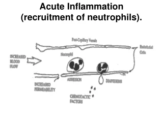
Leukocyte Cellular Events • Margination and Rolling • Adhesion and Transmigration • Migration into interstitial tissue
SLOWING CONCENTRATION Margination Rolling Adhesion Transmigration
Mucin-like glycoproteins (Sialyl-Lewis X PSL-1 & ESL-1) Integrins Selectins CAMS Weak and transient binding Results in rolling Integrins upregulated and activated for increased affinity to CAMS Results in firm adhesion
Margination • Normal flow - RBCs and WBCs flow in the center of the vessel • A cell poor plasma is flowing adjacent to endothelium • As blood flow slows, WBCs collect along the endothelium Margination
Endothelial Activation • The underlying stimulus causes release of mediators which activate the endothelium causing selectins and other mediators to be moved quickly to the surface
Selectins • Selectins bind selected sugars • Selected + Lectins (sugars) = Selectins • Some selectins are present on endothelial cells (E-Selectin) • Some selectins are present on leukocytes (L-Selectin) • Some selectins are present on platelets (P-Selectin) • Weak & transient binding • Results in rolling
Rolling • Selectins transiently bind to receptors • PMNs bounce or roll along Rolling
Adhesion • Mediated by integrins ICAM-1 and VCAM-1
Transmigration • Mediated/assisted by PECAM-1 & ICAM-1 (Integrins) • Diapedesis (cells crawling) • Primary in venules • Collagenases degrade BM Permeability
Chemotaxis • Movement toward the site of injury along a chemical gradient • Chemotactic factors include • Complement components (20 serum proteins) • Arachadonic acid (AA) metabolites • Soluble bacterial products • Chemokines, cytokines
Phagocytosis & Degranulation • Phagocytosis (engulf and destroy) • Degranulation and the oxidative burst destroy the engulfed particle • Recognition & attachment • Opsonins coat target and bind to leukocytes • Engulfment • Killing/degradation • O2 dep: Reactive O2 species in lysosomes & EC • O2 indep: Bactericidal permeability agents, lysozyme, MBP, lactoferrin
Leukocyte-induced tissue injury • Lysosomal enzymes are released into the extracellular space during phagocytosis causing cell injury and matrix degradation • Activated leukocytes release reactive oxygen species and products of arachidonic acid metabolism which can injure tissue and endothelial cells • These events underlie many human diseases (e.g. Rheumatoid arthritis)
Leukocyte adhesion deficiency 1 (LAD-1) • Recurrent bacterial infections • Inflammatory lesions lack neutrophil infiltrate • High numbers of neutrophils in the circulation • Neutrophils from patients can roll but do not stick • chain of CD11/CD18 integrin • Transfuse patients with normal neutrophils and they can emigrate
Mechanism of leukocyte adhesion deficiency 1 (LAD -1) • Absence of integrins on neutrophils • Mutation in n-terminal region of the integrin chain inhibits proper integrin assembly • Normal function is restored following transfection of patient cells with cDNA for chain
Chediak-Higashi Syndrome • This syndrome has been on every board test since Noah • Defect in chemotaxis and lysosomal degranulation into phagosomes
Chronic Granulomatous Disease • Defect in NADPH oxidase system • Marked decrease in ability to kill microorganisms
Chemical mediators of inflammation • Plasma-derived • Circulating precursors • Have to be activated • Cell-derived • Sequestered intracellular • Synthesized de novo • Most mediators bind to receptors on cell surface but some have direct enzymatic or toxic activity • Mediators are tightly regulated
Tissue injury Vasoactive mediators (eg. histamine) Chemotactic factors (eg. c5a) Production of inflammatory mediators Recruitment of inflammatory cells Increased vascular permeability Acute inflammation Chronic inflammation Edema PMNs Monos
Plasma Mediator Systems - Interaction 1. Kinin 2. Clotting 3. Complement 4. Fibrinolytic
Plasma Mediator Systems - Interaction Complement C5 XII C3 C5a C3a Kinin XIIa Prekallikrein Kallikrein High Mol. Wt. Kininogen Bradykinin Plasminogen Plasmin Fibrin FSPs Fibrinopeptides Prothrombin Thrombin Fibrinogen Fibrinolytic Clotting
Kinin cascade • Leads to formation of bradykinin • Bradykinin causes • Increased vascular permeability • Arteriolar dilatation • Smooth muscle contraction • Bradykinin is short lived (kininases) • Vascular actions similar to histamine
Complement system • Role in immunity (C5-9 complex) • Membrane Attack Complex (MAC C5-9) • Punches a hole in the membrane
Complement system • Role in inflammation (c3a and c5a) • Vascular effects • Increase vascular permeability and vasodilation • Similar to histamine • Activates lipoxygenase pathway of arachidonic acid metabolism (c5a)
Complement system • Leukocyte activation, adhesion and chemotaxis (c5a) • Phagocytosis • c3b acts as opsonin and promotes phagocytosis by cells bearing receptors for c3b
Inflammatory Mediators from Complement Anaphylatoxins: C3a, C5a, & C4a trigger mast cells to release histamine and cause vasodilatation C5a also activates the lipoxygenase system in PMNs and monocytes release of inflammatory mediators Leukocyte activation, adhesion, & chemotaxis: C5a activates leukocytes, promotes leukocyte binding to endothelium via integrins and is chemotactic for PMNs, monos, eos, & basos