Download
1 / 50
540 likes | 1.05k Views
Endocrine glands. Morphology and embryology Course: Endocrine system David Kachlik and Petr Zach. Endocrine glands Glandulae endocrinae. One of regulatory systems hormone (greek horman – to awake, stimulate)

E N D
Endocrine glands Morphology and embryology Course: Endocrine system David Kachlik and Petr Zach
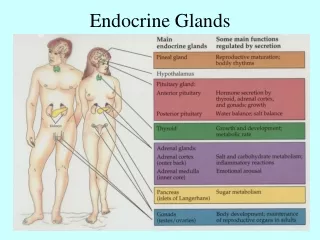
Endocrine glandsGlandulae endocrinae • One of regulatory systems • hormone (greek horman – to awake, stimulate) • Chemical messenger produced by endocrine gland and transported by blood to target organs • proteins (polypeptides) - insulin • Biogennic amines - adrenalin • steroids - estrogens
Endocrine glandsHistory • Thomas Wharton • 1614-1673 • 'Adenographia‚ • First detailed description of endocrine glands
Endocrine glandsHistory • Ernest Henry Starling • 1866-1927 • Created general rules of „internal secretion “ • He used already existing word „hormones“ • Sir William Bate Hardy, physiologist from Cambridge
Endocrine glandsOrganization • glands • Disseminated cells • neuroendocrine cells
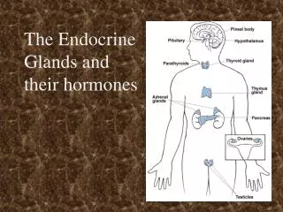
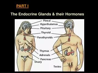
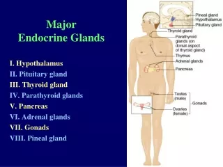
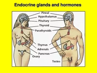
Endocrine glandsGlands • Hypothalamus • Hypophysis; Gl. pituitaria • Glandula thyroidea, thyroid gland • Gll. parathyroideales • Gll. Suprarenales, suprarenal gland • Langerhans islets (Insulae pancreaticae) • Gl. pinealis; Corpus pineale
Hypothalamus + hypophysis Systema hypothalamo-hypophysiale Hypotalamo-hypophyseal axis
Hypophysis; Glandula pituitariaHistory • Galén – mucus produced for nasal mucosa • Schneider – 1655 rejects Galén theory • Minkowski, Hutchinson – connection between growth problems and hypertrophy of hypophysis • Cushing – explained function of hypophysis „dirigent of endocrine system, minister head“
Hypothalamus • Bazal part of diencephalon (Diencephalon), bazally to IIIrd ventricle • Function • Gathers info from body and surroundings • Highest autonomic center (vegetative) • Part of limbic system • Directs other endocrine glands • Corpora mamillaria, tuber cinereum, infundibulum, hypophysis
Hypothalamus • Anterior hypothalamus – ncl. magnocellularis • Ncl. paraventricularis + supraopticus – oxytocin and vasopressin (ADH) • Middle hypothalamus (tuber cinereum) – ncl. parvocelularis • Ncl. arcuatus – regulation of adenohypophysis • Posterior hypothalamus
Hypothalamus - Hormones • Ncl. arcuatus - production • Eminetia mediana – release into first capillary bedstream • Releasing hormones (releasing) • SRH, PRH, GnRH, TRH, CRH • Supressing hormones (inhibiting) • somatostatin, PIH (= dopamin)
Hypophysis - anatomy • Formed by two lobes • ventral - adenohypophysis • dorsal – neurohypophysis • Located in sella turcica ossis sphenoidalis • Covered by dura mater – diaphragma sellae • foramen diaphragmatis Pacchioni – for hypophyseal infundibulum • Transsphenoid approach
Hypophysis - anatomy • Ventral lobe (Adenohypophysis; Lobus anterior) • Pars distalis (principalis) – biggest • Pars intermedia – between both lobes • Pars tuberalis – part by infundibulum • Posterior lobe (Neurohypohysis; Lobus posterior) • Lobus nervosus (Pars nervosa) – proper posterior lobe • Infundibulum – stalk leading to hypotalamus
Blood supply of hypophysis • Hypophyseal portal system • A. hypophysialis inferior (from pars cavernosa to neurohypophysis) • A. hypophysialis superior (from pars cerebralis via hypothalamus into adenohypophysis) • Vv. hypophysiales into sinus cavernosus
Hypophysis - development • Ratkhe pouch (from ectoderm) • 3rd week – in the roof of mouth cavity • Exagination towards diencephalon • Separation of exagination, proliferation of ventral wall • Exagination of basis of diencephalon • Creates posterior lobe • Differentiation into pituicytes (glia)
Ventral lobe = Adenohypophysis • Pars distalis • biggest (75%) • Pars tuberalis • cranially • Pars intermedia • between adeno- and neurohypophysis
Pars distalis adenohypophysis • Strand of cells (chordae endocrinocytorum), between them capillaries • on HE 3 types of cells • acidophilic • bazophilic • PAS-pozitive • chromophobic • w/o granules, non-differentiated elements
Pars distalis – Acidophilic cells • α – cells (Endocrinocytus somatotropicus) • Rough granules, GER • Around nucleus zone w/o granules - GA • Growth hormone (somatotropin, GH) • ε – cells (Endocrinocytus prolactinicus) • Usually small, not numerous (x gravidity, lactation) • Small granules (enlargement in gravidity) • prolaktin (PRL)
Pars distalis – bazophilic cells • β1 – cells (Endocrinocytus corticotropicus) • Big granules close to cell membrane • ACTH, β-MSH, Met-enkefalin, endorfin • β2 – cells (Endocrinocytus thyrotropicus) • Large cells, small granules by BM • TSH • δ – cells (Endocrinocytus gonadotropicus) • Large cells, middle size granules • FSH, LH
Adenohypophysis electron microscope modifikovaný Azan imunoperoxidase reaction to LH • HE
Pars tuberalis adenohypophysis • surrounds infundibulum • Numerous capillaries • mostly δ – cells • Little bit β2 – cells
Pars intermedia adenohyophysis • rudimentary • Cells compose celumns • Bazophilic cells • Could be present sac (Ratkhe´s folliculus)
Posterior lobe = Neurohypophysis • Eminentia mediana • Bottom of IIIrd ventricle • Numerous non-myelinated nerve fibers • stalk = Infundibulum • Tractus hypothalamohypophysialis • Neurofibra neurosecretoria (+ vesicula neurosecretoria) – non myelinated nerve fibers) • Some terminate by capillaries • Lobus nervosus; Pars nervosa
Lobus nervosus / Pars nervosa neurohypophysis • Nerve fibers • Axons of neurons from hypotalamus • Corpusculum neurosecretorium (Herring corpuscles) – accumulation of granules • oxytocin + ADH (antidiuretic hormone = vasopressin) • pituicytes • Glial cells • capillaries (synapsis neurohaemalis)
Examination and diseases • CT • Levels of hormones • Tumors of hypophysis – usually benign, hormally active • Sheehan syndrome – after delivery bleeding into hypophysis
Thyroid gland – history • Galén – to water inside of pharynx • Paracelsus – struma + cretenism • Wharton 1614-1673 – to make neck more beautiful • 1844 Simon – endocrine gland • 1891 Murray – administration of extract from TG • 1895 Baumann – TG contains iodine components
Thyroid gland - anatomy • Glandula thyroidea • thyroxin T4, trijodtyronin T3 • kalcitonin • Located at the level C6-C7 • 2 lobes – lobus dexter + sinister • isthmus (on 2nd-4th tracheal cartillage) • capsula fibrosa – 2 sheets – stroma • parenchyma + lobuli
Thyroid gland – arteries and veins • A. thyroidea superior (from a. carotis externa) • A. thyroidea inferior (z truncus thyrocervicalis) – crossing with n. laryngeus recurrens • A. thyroidea ima Neubaueri (from arcus aortae) - 2 % • Vv. thyroideae superiores et mediae Lichačeva (into V. jugularis interna) • Plexus thyroideus impar (into v. brachiocephalica sinistra)
Thyroid gland - development • Development since 24th day • Exagination of endoderm of primitive pharynx • Relative and absolute descencus - ductus thyroglossus • foramen caecum • gll. thyroideae accessoriae • Lobes origin • lobus pyramidalis • ligamentum suspensorium gl. thyroideae / musculus levator glandulae thyroideae (smooth)
Thyroid gland - histogenesis • solid endoderm formation • Ingrowth of surrounding mezenchyme and blood vessels • Ingrowth of ultimobranchial corpuscles • 10th week – separation of cells into groups • Single layer epithelium around lumina • 11th week – production of koloid
Thyroid gland - composition • capsule (capsula fibrosa) • stroma - septum (septas between lobes) • lobus lobulus folliculus • follicles (50 - 900 μm) • spherical • Single layer epithelium of follicular cells • contains colloidum (coloid) - thyroglobulin • Follicular cells (thyrocytus T) • Parafollicular cells (thyrocytus C)
Follicular cells (Thyrocytus T) • Spherical nucleus • numerous gER (bazally) and MIT • Numerous lyzosomes • thyroglobulin, cleavage of T4 and T3
1. iodine pump using ATP recirculates iodine from blood into colloid 2. and 3. synthesis of thyroglobulin and peroxidase, deposition in one secretory vesicle and its release into colloid by exocytosis 4. iodination off thyroglobulin using peroxidase in colloid and formation of iodinethyroglobulin Endocytosis of iodinethyroglobulin 5. fusion of primary lyzozome with this vesicle Proteolysis of iodinethyroglobulin into T3, T4 and other fragments Release of T3 and T4 into circulation 6. binding of transport plasmatic protein Synthesis of hormones of thyroid gland
Parafolicular cells (thyrocytus C) • C-cells • Located between follicles (individually, also goups) • Bigger, lighter • numerous gER as well as GA, MIT • granules – spherical, dark • Prooduction and accumulation of kalcitonin
Thyroid gland – examination • ultrasound • Scintigraphy with radioactive iodine 131
Thyroid gland – disease • ultrasound • scintigraphy with radioactive iodine 131 • Less then 10μg iodine per day struma from lack of iodine • hypotyroidizmus • kretenizmus (children) – screening in newborns • Myxedema (adults) • autoimmune – Hashimoto struma • hypertytoidizmus (tyreotoxicosis) • autoimmune – exophtalmic struma = Graves-Basedow disease
Parathyroid gland - characteristic • Gll. parathyroidea superior et inferior • 2 pairs of small spheroid structures on the posterior side of lobes of thyroid gland • Individual branches from a. thyroidea inferior • Role in bone metabolism • parathormon (PTH)
Parathyroid gland - development • Dorsal part of IIIrd and IVth branchial exagination • 5th week – proliferation of endoderm, loss of lumen • Ingrowth of vessels from mesenchyme • Principal cells – fetal metabolism of calcium • Oxyphilic cells – origin in 7th year of life
Parathyroid gland - composition • capsule + septas • parenchyme parcellated into columns • Principal cells (parathyrocytus endocrinus) • Relatively large (4-8 um) • light cytoplasm • Granules containing PTH • Oxyphilic cells (Parathyrocytusoxyphilicus) • Rare, bigger • cytoplazm darker, w/o granules, numerous MIT • Function not clear
Parathyroid gland disease • hyperparatyroidizmus • primary (adenoma) • Patological calcification of tissue (based on hypercalcemia) • Recklinghausen bony osteodystrofia (fractures) • nephrolitiasis • secondary (reactive hyperplazia PT in hypocalcemia in kidney diseases) • terciary (in case of successful kidney transplantation)
Parathyroid gland - disease • hypoparatyroidizmus • tetany • iatrogenic in case of tumor excision • Transplantation of gland on the forearm subcutaneously • examination – nuclear medicine