Download

1 / 56
620 likes | 989 Views
ESOPHAGEAL CARCINONA. Anatomy Of Esophagus. Anatomy Of Esophagus. Anatomy Of Esophagus. Anatomy Of Esophagus. Histology Of Esophagus. Esophageal Cancer.

E N D
Esophageal Cancer Esophageal cancer is the seventh leading cause of cancer death worldwide. Incidence of esophageal carcinoma can be as high as 30-800 cases per 100,000 persons in particular areas of northern China, some areas of southern Russia, and northern Iran. Unlike in the United States, squamous cell carcinoma is responsible for 95% of all esophageal cancers worldwide.
Esophageal Cancer Esophageal cancer is generally more common in men than in women, with a male-to-female ratio of 3-4:1. Esophageal cancer occurs most commonly during the sixth and seventh decades of life. The disease becomes more common with advancing age; it is about 20 times more common in those older than 65 years than in persons younger than 65 years.
Risk Factors A- Dietary Factors 1-deficiency of Vitamins(A,C,B1,2,6) 2-deficiency of trace elements(Zinc) 3-Fungal contamination of foodstuffs 4-High contents of nitrites/nitrosamines B-Lifestyle 1-burning hot beverages or foods 2-Alcohol consumption 3-Tobacco use
Risk Factors C- Precancerous conditions 1-Barrett Esophagus 2-Tylosis palmaris 3-Achalasia cardia 4-Plummer-Vinson Syndrome 5-Caustic Strictures D- Genetic Predisposition 1-Ectodermal dysplasia (Ectodermal dysplasia is a group of conditions in which there is abnormal development of the skin, hair, nails, teeth, or sweat glands.)
Risk Factors 2- Racial Predisposition • Areas of northern China ( Linxian in Henan province ), some areas of southern Russia, and northern Iran. 3-Epidermolysis bullosa (Epidermolysisbullosa (EB) is a group of inherited bullous disorders characterized by blister formation in response to mechanical trauma).
Types Of Cancer • Squamous cell Carcinoma ( cigarette smoking and alcohol ) • Adenocarcinoma ( GERD )
Types Of Cancer By far the commonest type is Squamous cell carcinoma comprising about 90-95% of all esophageal carcinomas outside united States. In US Adenocarcinoma contribute to about 50% of all esophageal carcinomas. Incidence of Adenocarcinoma is increasing throughout the world with a concomitant increase in the incidence of GERD, the link being the Barrett esophagus.
Squamous Cell Carcinoma Sites 50% in the Middle 1/3 20% in the upper 1/3 30% in the lower 1/3 Patterns 1-polypoid exophytic lesions- 60% 2-diffuse infiltrative type - 15% 3-Excavated(Ulcerated type) - 25%
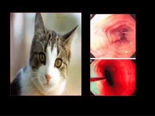
Clinical Features 1- Dysphagia The most common presenting symptom.Dysphagia is initially experienced for solids, but eventually it progresses to include liquids. A complaint of dysphagia in an adult should always prompt an endoscopy to help rule out the presence of esophageal cancer. A barium swallow study is also indicated. 2- Weight loss is the second most common symptom and occurs in more than 50% of people with esophageal carcinoma
Clinical Features 3- Pain can be felt in the epigastric or retrosternal area. It can also be felt over bony structures, representing a sign of metastatic disease. 4- Hoarseness caused by invasion of the recurrent laryngeal nerve is a sign of unresectability. Patients may have a persisting cough. 5- Respiratory symptoms can be caused by aspiration of undigested food or by direct invasion of the tracheobronchial tree by the tumor. The latter is also a sign of unresectability.
Physical Finings The examination findings are often normal. Emaciation i-e weight loss and weakness. Pallor-due to chronic anemia due to bleeding Hepatomegaly may result from hepatic metastases. Lymphadenopathy in the laterocervical or supraclavicular( Troisier’s sign ) areas represents metastasis and, if confirmed by needle aspiration or biopsy findings, is a contraindication to surgery.
Spread 1- Local or Direct spread The cancer starts as mucosal ulceration which spreads to submucosa. The spread occurs transversely and longitudinally. Once it invades all the layers, the structures in the vicinity are involved. Trachea- Tracheo-esophageal fistula develops from invasion of trachea in case of carcinoma upper 1/3 . Bronchi- Trancheo-bronchial fistula from carcinoma middle 1/3. Aorta- Esophago-aortic fistula Results in massive bleeding( one of the cause of death ).
Spread 2- Lymphatic Cervical part drains into Left and Right Supraclavicular nodes . Thoracic part drain into Tracheo-esophageal Para-esophageal nodes. Abdominal part drain into Nodes along the lesser curvature and then into celiac nodes. Accordingly Lymphatic spread may occur into these nodes according to the site of involvement of esophagus.
Spread 3- Hematogenous It results in secondary deposits in Liver which clinically appears as Nodular Enlarged Liver. Later, Ascitis and recto-vesical deposits occur.
Investigations 1- CBC may show Anemia 2- LFTs – if secondaries in liver occur –Increased ALP 3- USG abdomen done to r/o liver secondaries, lymph nodes in the porta hepatis, celiac nodes etc. 4- Barium swallow demonstrates irregular persistent intrinsic filling defects. 5- esophagoscopy to visualize the growth and to take biopsy. 6- Multiple biopsies- in high risk areas like China Endoscopic staining with supravital dyes (indigocarmine)is done 2 identify dysplasticepithelium
Investigations 7- CXR to r/o aspiration pneumonia and mediastinal widening and posterior tracheal indentation. 8- Bronchoscopy to r/o involvement of bronchus. 9- CT-Scan of chest to find out local infilteration. It is very useful before doing esophagectomy to asses the vital structures involvement like bronchus,airway etc. 10- Endoscopic Ultrasound to know the depth of the wall involvement,to detect mediastinal lymph nodes etc.
Treatment Non-Surgical Majority of esophageal cancers are advanced at the time of diagnosis. In such situation the goal of treatment is Palliation of Dysphagia allowing the patients to eat. Various modalities are available. 1- Radiotherapy successful in relieving dysphagia in about 50% of cases combined with chemo.
2- Laser therapy Can help achieve temporary relief of dysphagia in as many as 70% of patients. Multiple sessions are usually required. 3- Chemotherapy Chemotherapeuting agents with promising response are Cisplatin,5-FU and Paclitaxel. 4- Photodynamic therapy offer an interesting non-surgical form of therapy.Activation of photosensitizer by Light in the desired area which causes biologic damage to abnormal tissue.
5- Metallic self expandable stents are currently the choice of tubes to relieve Dysphagia.
Surgical Treatment Esophageal resection (esophagectomy) remains a crucial part of the treatment of esophageal cancer. It is used in patients who are considered candidates for surgery. An esophagectomy can be performed by using an abdominal and a cervical incision with blunt mediastinal dissection through the esophageal hiatus i-e transhiatal esophagectomy[THE] or by using an abdominal and a right thoracic incision i-e transthoracic esophagectomy[TTE].
T-H-E Trans-haital esophagectomy is usually done in a mobile growth of Lower 1/3 of Esophagus without Thoracotomy. Esophagus and part of stomach and the involved lymph nodes are removed F/B Gastric pull-up And anastomosis in neck.