Download

1 / 28
540 likes | 2.27k Views
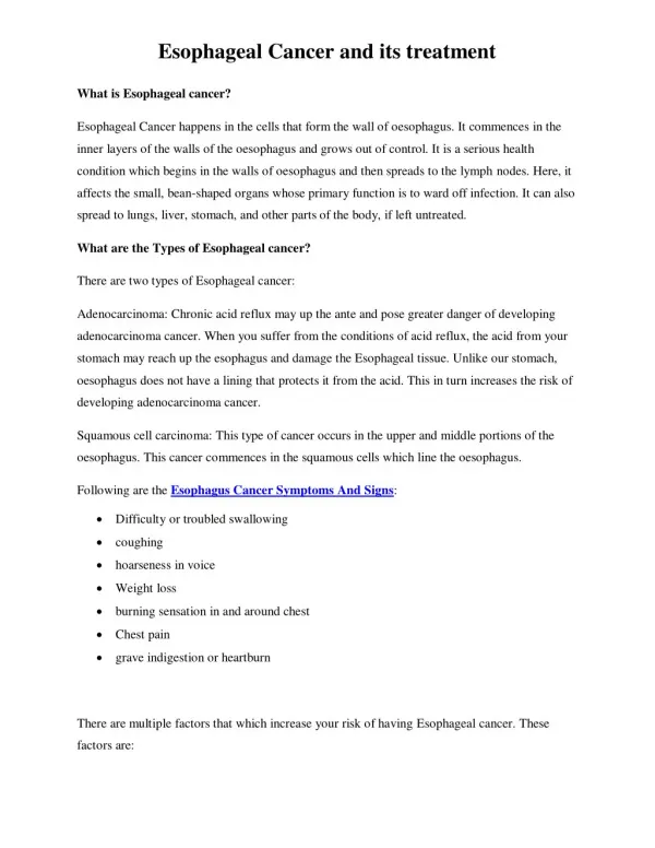
ESOPHAGEAL CANCER. KHANI.M,MD MEDICAL ONCOLOGIST&HEMATOLOGIST ISFAHAN BLOOD&CANCER INSTITUE WWW.IBCI.IR. First step?. CASE NO :1. 55 Y old previously obese man Progressive dysphagia & chest pain & weight loss since 3 month ago Past history of GERD for 5 years+smoking 20p/y

E N D
ESOPHAGEAL CANCER KHANI.M,MD MEDICAL ONCOLOGIST&HEMATOLOGIST ISFAHAN BLOOD&CANCER INSTITUE WWW.IBCI.IR
CASE NO :1 • 55 Y old previously obese man • Progressive dysphagia & chest pain & weight loss since 3 month ago • Past history of GERD for 5 years+smoking 20p/y • PHE:cachexia,temporal muscle wasting,no lap • LAB:hb=10,plt=180,000,serum alb=2.5
Endoscopy result • A polypoid 3*3 cm mass lesion in distal esophageus near z line,multiple biopsy taken the scope don’t pass the lesion so evaluaion of stomach wasn’t possible
EUS wasn’t available • Thoracic ct scan:mass like lesion in distal esohagus highly suggest GE junction tumor, regional(diaphragmatic&pericardial) lap present • Echo=EF 65%.mild pericardial effusion
Abdominoplvic ct scan: no lymphadenopathy, no metastatic lesion
Laparascopy? • PET • PET/CT • Pelvic &cervical ct?
Pathology report • Poorly differentiated adenocarcinoma • Tumor invades muscularispropria
Clinical staging • T2,N1,M0 • Stage IIb
Next step? • Surgery • Radiation • Chemoradiation then surgery • Definitive chemoradiation • Chemo then chemorad then surgery +- chemo
This patient recived 2course ECF then chemoradiation then surgery
SURGERY • TRANSHIATAL
Post surgery pathology report • Poorly differentiated adenocarcinoma • All margin free • Tumor invades adventitia • 4 out of 4 lymph node free of tumor • T3N0/1M0 at least IIA
NEXT? • 2 more ECF • Follow up
CASE NO:2 • A 60 y/o male • PMH:CLL BINET B (chlorambucil+pred),SCC of face skin (surgery+radiation+flap),DM(insulin) • Heavy smoker, • Progressive dysphagia&weight loss since 3 mo ago+intractable vomiting • PHE:several deformity of face due to flap with dirty wound,cervical lap 2*3 cm,hoarsness
ENDOSCOPY RESULT • A MASS LESION LOCATED IN 25 CM OF INCISURA TEETH CAUSE NEAR COMPLETE OBSTRUCTION OF LUMEN,MULTIPLE BIOPSY WAS TAKEN
PATHOLOGY REPORT • SQUAMOUS CELL CARCINOMA
NEXT STEP? • CERVICAL,THORACIC CT SCAN • ABDOMINOPELVIC CT? • BRONCHOSCOPY • LAPAROSCOPY? • PET OR PET/CT? • EUS
RESULTS • MASS LESION IN THORACIC ESOPHAGUS,DIMINISH FAT STRIP BETWEEN TRACHEA&ESOPHAGUS WITH MULTIPLE MEDIASTINAL LAP • NORMAL ABDOMINOPELVIC CT • BRONCHOSCOPY:NORMAL,NEGATIVE BIOPSY OF SUSPICIOUS AREA
T3/T4,N1,M0 • STAGE III
NEXT STEP? • SURGERY • DEFINITIVE CRT • CX -> CRT-> CX ->+-SURGERY • CX-> CRT > SURGERY
AFTER 5 WEEK • CONTINUE DYSPHAGIA • ENDOSCOPY:DECREASED STENOSIS BUT PERSISTANT MASS • THORACIC CT:NO OBVIOUS CHANGES COMPARE TO PREVIOUS CT,NO OBVIOUS OTHER ORGAN ADHESION
PET • BIOPSY
RADIATION • SURGERY • CX