Download
1 / 39
470 likes | 938 Views
ESOPHAGEAL TUMORS. Aswad H. Al.Obeidy FICMS, FICMS GE&Hep Kirkuk General Hospital. Eso tumors. Malignant > common than benign. Unfortunately, eso cancer often discovered late & overall 5 y ear prognosis is bad < 10%. Even for potentally resectable ca eso, 5 y survival is < 30%.

E N D
ESOPHAGEAL TUMORS Aswad H. Al.Obeidy FICMS, FICMS GE&Hep Kirkuk General Hospital
Eso tumors • Malignant > common than benign. • Unfortunately, eso cancer often discovered late & overall 5 y ear prognosis is bad < 10%. • Even for potentally resectable ca eso, 5 y survival is < 30%
Benign Neoplasms • The most common is a gastrointestinal stromal tumour (GIST, another name for leimymoma),usually asymptomatic but may cause bleeding or dysphagia • Others, include fibrovascular polyps, papillomas, lipomas, neurofibromas, granular cell tumors. • When large, can cause dysphagia or chest pain from obstruction or stretch. • Usually discovered incidentally.
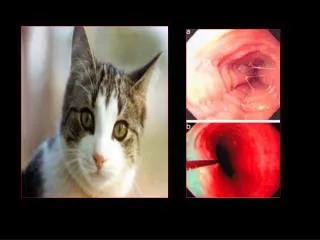
LEIOMYOMA OF OESOPHAGUS • Most common benign tumor of esophagus & small bowel but not common in the colon • Usually asymptomatic • May produce dysphagia or hematemesis if large. • Typically occurs in young males • Found most often in distal third of esophagus. • Usually solitary, but may be multiple (3%). • Imaging findings: • Smooth, sharply-marginated mass. • Well-defined, intramural (wall) mass &may narrow the lumen. • May have coarse calcifications (only calcifying esophageal tumor) • Rarely ulcerates
LEIOMYOMA OF OESOPHAGUS: DIAGNOSIS • Barium swallow. • Endoscopy: smooth submucosal lesion.
ETIOLOGY & PATHOGENESIS • Almost all are adenocarcinoma or squamous cancers. • Small-cell cancer is a rare third type.
SCC • In West relatively rare (4 cases /100 000). • Common in Iran, Iraq, Africa , China, (200/100 000). • Can arise in any part of the oesophagus from the post-cricoid region to the cardia. • Almost all tumours above the lower third of the oesophagus are squamous cancers.
Adeno ca • Arises in the lower third of the oesophagus from Barrett's oesophagus or from the cardia of the stomach. • The incidence is increasing & now 5:100 000 in UK; possibly because of the high prevalence of GERD/ Barrett's.
ETIOLOGY & PATHOGENESIS • Overall taking both types, no sex difference. • SCC > in women. • Relatively common in Kurdish. • Should be considered in any case presenting with dysphagia.
SCC:Risk factors • Alcohol • Tobacco smoking • SCC of the head & neck • Lye or post-caustic strictures • Achalasia • Papilloma virus infection • Plummer-Vinson syndrome • Tylosis (familial hyperkeratosis of palms & soles) • Celiac disease • Radiation exposure • Post-cricoid web
SYMPTOMS • The most common is progressive dysphagia over a several-month period until only liquids can be taken. • The obstruction does not occur until the cancer is far advanced. • The dysphagia may be accompanied by a steady, boring pain, which often signals mediastinal involvement & inoperability.
SYMPTOMS • Unexplained persistent chest pain should always be investigated by a careful double-contrast Barium or endoscopy. • More advanced; halitosis & weight loss. • Coughing after drinking fluid may be caused either by nearly complete esophageal lumen obstruction, with overspill into the larynx, or by the development of a tracheoesophageal fistula. • Hematemesis & Hoarseness from involvement of the recurrent laryngeal nerve by tumor are unusual symptoms.
SIGNS • Weight loss. • Nail bed clubbing can be seen with both benign & malignant tumors. • Vricho’s node in left supracalvicular region. • Early diagnosis affords the only chance for cure.
DIAGNOSIS • The investigation of choice is upper GI endoscopy with cytology & biopsy, always a good retroflexed view of the cardia from below, to make certain that an adenocarcinoma in GEJ has not been overlooked • A barium swallow demonstrates the site& length of the stricture but adds little useful information. • Once a diagnosis has been achieved, investigations are performed to stage the tumour& define operability. • Thoracic & abdominal CT are carried out to identify metastatic spread & local invasion. • Invasion of the aorta&other local structures may preclude surgery. • Unfortunately, CT understages tumours &the most sensitive modality is EUS to define the TNM stage.
DIAGNOSIS • Biopsy of visible tissue may reveal only inflammation; so as many as 6-9 deep biopsy specimens should be obtained.
DIAGNOSIS: STAGING • Evaluation for local tumor spread, mediastinal nodal involvement & liver metastases is essential for staging before a therapeutic decision is reached by: • Physical examination for lymphadenopathy • Tests of liver enzymes • Chest radiography • CT scan • For upper & mid-esophageal tumors, bronchoscopy is indicated to evaluate for asymptomatic invasion of the tracheobronchial tree • Endoscopic ultrasound (EUS) is useful to detect the level of invasion & presence of mediastinal lymph node abnormalities & is becoming the favored test to determine if a lesion is resectable
The tumour (T) has extended through oeso wall (stage T3). A small peri-tumoral lymph node (LN) is also seen.
TREATMENT • Choice of therapy depends on: • Location • Size • Presence or absence of spread • Cell type
TREATMENT • Endoscopic mucosal dissection or resecion is an option for early EC, a rare situation.
TREATMENT • Surgical resection of SCC & adenocarcinoma of the lower 1/3 is preferred unless widespread metastases present. • Surgery offers the benefit of rapidly restoring esophagogastric continuity. • Only 1/4 have a resectable tumor; of these, 10 - 20% do not survive the operative period. • 5-year survival is only 5 - 20%, even with extensive resection. • Long-term survival cannot be predicted in the individual case by the operative findings. • There is growing enthusiasm for palliative resection with restoration of GI continuity with stomach or colon.
TREATMENT • Esophagectomy still remains the standard modality to cure otherwise fit patients with early stage& surgical mortality continues to improve in high-volume centers, from 1% to 5%. • For some, with cardiopulmonary or hepatic morbidities may be treated endoscopically with local excision, thermal or nonthermal laser, or cryoablation. • Local endoscopic therapy significantly prolonged survival in high-risk patients with clinical T0 or T1, N0 EC& is a reasonable alternative for those patients who are not candidates for potentially curative esophagectomy.
TREATMENT • When obvious extraesophageal spread is present, palliation may be achieved with dilation+/- Endoscopic metalic stenting to restore & maintain an adequate esophageal lumen.
TREATMENT • Destruction of intraluminal tumor & restoration of an adequate lumen may be performed by endoscopic laser therapy, intraluminal heat-coagulating probe, or photodynamic therapy.
TREATMENT • Despite modern treatment, the overall 5-year survival of oesophageal cancer is 6-9%. • Survival following oesophageal resection depends on stage. • Tumours which have extended beyond the wall,have lymph node involvement (T3, N1) are associated with a 5-year survival of around 10% after surgery. • Without LNs, Overall survival following 'potentially curative' surgery (all macroscopic tumour removed) is about 30% at 5 years& can be improved by neoadjuvant (pre-operative) chemotherapy with agents as cisplatin/ 5-fluorouracil.
ADENOCARCINOMA • A rapid rise in adenocarcinoma, particularly in white men, has made their current cancer rates about equal or even higher in the west. • Unlike SCC, arise in the distal esophagus because of the presence of Barrett’s eso, a complication of GERD. • Lymphatic spread is common. • Adenocarcinomas are radio insensitive; although chemoradiation &surgery may improve survival, the 5-year survival < 10% almost equal to SCC. • Palliation is the same as for inoperable SCC.