Download
1 / 85
960 likes | 1.49k Views
Dr Stephen O'Hanlon MA(Oxon) MBBChir(Cantab) MRCS DOHNS MSc. Examination of the Ears, Nose, Throat, and Neck. Curriculum statement 15.4 ENT and Facial Problems. Examination of the Ears, Nose, and Throat. 7) Otoscopic examination 8) Fistula test 9) Free field testing 10) Tuning fork tests

E N D
Dr Stephen O'Hanlon MA(Oxon) MBBChir(Cantab) MRCS DOHNS MSc Examination of the Ears, Nose, Throat, and Neck
Curriculum statement 15.4 ENT and Facial Problems Examination of the Ears, Nose, and Throat
7) Otoscopic examination 8) Fistula test 9) Free field testing 10) Tuning fork tests 11) Facial nerve 12) Post nasal space Examination of the Ear • 1) Introduction • 2) Position the patient • 3) Start with the better ear • 4) Inspect the pinna • 5) Inspect the mastoid • 6) Inspect the external auditory meatus
Examination of the Ear • 1) Introduce yourself to the patient • Any deafness? • Communication
Examination of the Ear • 2) Position the patient • Away from the wall • In chair • Can walk around patient
Examination of the Ear • 3) Start with the better side • Start with the dry side • Remove hearing aids
Examination of the Ear • 4) Inspect the pinna • Front and behind • Skin condition • Lesions • Scars • Pre-auricular area (common place for sinus) • Condition of cartilage
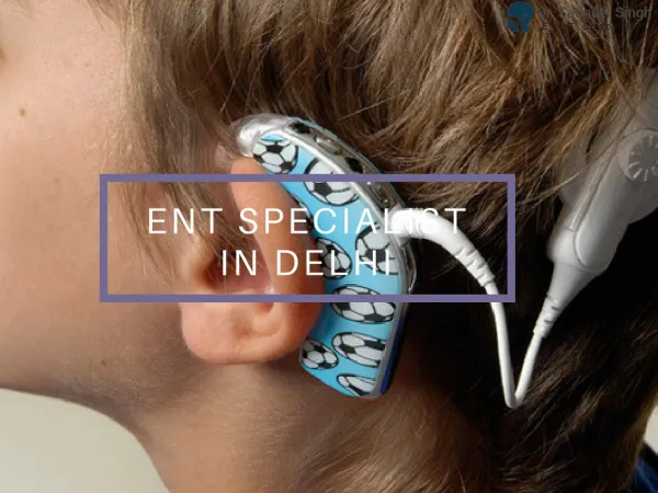
Examination of the Ear • 5) Inspect the mastoid • Mastoiditis is very, very rare • Post auricular lymph nodes • Scars • BAHAs and Cochlear implants
Examination of the Ear • 6) Inspect the external auditory meatus • Pull pinna upwards, outwards and backwards • In infants downwards and backwards • In children pull backwards • Otorrhoea and otomycosis • Canal stenosis • Exostoses and osteomas
Examination of the Ear • 7) Otoscopic examination • The lateral process and handle of the malleus lie towards the centre of the tympanic membrane • Four quadrants • Perforation • Central or marginal • What can be seen through it • Mastoid cavity • Dry • Wet, inflamed
Examination of the Ear • 8) Fistula test • A test for ENT doctors! • Warn the patient • A cholesteatoma has erroded part of a semi-circular canal • Pressure in the EAM causes conjugate deviation of the eyes
Examination of the Ear • 9) Free field testing • Start with the better ear • Use masking, such as a tragal rub • Whispered voice, conversation voice, and shouted voice at 60cm • Use double number fingers or bisyllable words
Examination of the Ear • 10) Tuning fork tests • Traditionally 512Hz (256Hz may be better) • Rinne and Weber (they were both German) • Help differentiate between conductive and sensorineual hearing loss • Limited reliability (around 80%) • For Weber, incisors > vertex > forehead
Examination of the Ear • 11) Facial nerve • Otoneurological conditions affect the cranial nerves closest to the vestibulocochlear nerve • Cholesteatoma • Malignant otitis externa (osteomyelitis) • Vestibular Schwannoma (formerly acoustic neuroma) • Other skull-base tumours
Examination of the Ear • 12) Post nasal space • Conditions affecting the middle ear can sometimes affect the post nasal space • Hopkins rod, flexible endoscope, or angled mirror usually needed • Massive lesions can sometimes be seen through the nose, or behind the soft palate
5) Anterior rhinoscopy 6) Oral examination 7) Post nasal space examination 8) Neck examination Examination of the nose • 1) Introduce yourself • 2) Position patient • 3) Inspect the external nose • 4) Inspect the nasal tip, vestibule, and nasal airways
Examination of the nose • 1) Introduce yourself • Any hyponasal speech?
Examination of the nose • 2) Position the patient • Head-mirror or headlight?
Examination of the nose • 3) Inspect the external nose • Compare nose to rest of face • Size and shape • Skin • Swelling, bruising, ulcers • Profile
Examination of the nose • 4) Examine the nasal tip, vestibule, and assess the nasal airways • Nasal tip • Nostrils and air flow • Mist test • Condition of mucosa inside vestibule
Examination of the nose • 5) Anterior rhinoscopy • Thudichum’s speculum vs otoscope • Obvious lesions • Mucosa • Septum • Turbinates (and osteomeatal complex)