Download
1 / 22
340 likes | 667 Views
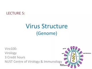
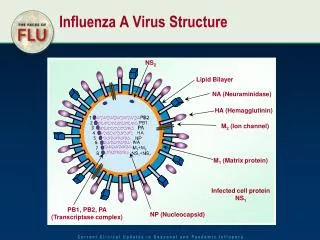
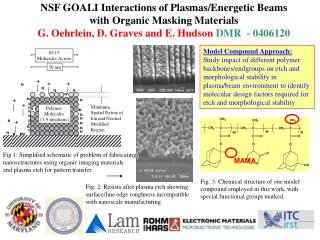
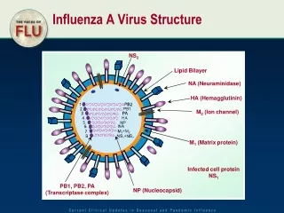
HA hemagglutinin protein - responsible for binding the virus to the cell that is being infected. Primary antigen for flu vaccines. Fig. 1. Schematic representation of influenza A virus genome and proteins.

E N D
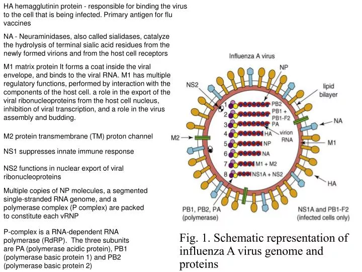
HA hemagglutinin protein - responsible for binding the virus to the cell that is being infected. Primary antigen for flu vaccines Fig. 1. Schematic representation of influenza A virus genome and proteins NA - Neuraminidases, also called sialidases, catalyze the hydrolysis of terminal sialic acid residues from the newly formed virions and from the host cell receptors M1 matrix protein It forms a coat inside the viral envelope, and binds to the viral RNA. M1 has multiple regulatory functions, performed by interaction with the components of the host cell. a role in the export of the viral ribonucleoproteins from the host cell nucleus, inhibition of viral transcription, and a role in the virus assembly and budding. M2 protein transmembrane (TM) proton channel NS1 suppresses innate immune response NS2 functions in nuclear export of viral ribonucleoproteins Multiple copies of NP molecules, a segmented single-stranded RNA genome, and a polymerase complex (P complex) are packed to constitute each vRNP P-complex is a RNA-dependent RNA polymerase (RdRP). The three subunits are PA (polymerase acidic protein), PB1 (polymerase basic protein 1) and PB2 (polymerase basic protein 2)
281 145 103 UBE1L 1 93 ISG15 186 223-237 PABII CPSF 73 1 NonStructural Proteins of Influenza Virus 1918-19 Flu Pandemic - > 20,000,000 deaths Flu A (Bird Flu) 50% mortality rate since 1997 dsRNA NS1B (32 kDa x 2): Human Flu dsRNA NS1A (27 kDa x 2): Bird Flu
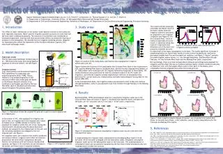
Schematic diagram of the multiple functions of NS1 within infected cells (a) Pre-transcriptional limitation of IFN-b induction. (b) Inhibition of the antiviral properties of PKR and OAS/RNase L. (c) Post-transcriptional block to processing and nuclear export of all cellular mRNAs. (d) Enhancement of viral mRNA translation. (e) Activation of PI3K. Translation initiation Hale et al. Journal of General Virology (2008), 89, 2359–2376
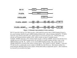
Sequence alignment of NS1A proteins from 5 different influenza strains referred to in this proposal
Solution NMR Structure of the dsRNA-binding domain of non-structural protein 1 of H5N1 bird virus (NS1A) R38: essential for dsRNA binding in vitro and viral virulence R38 R38’ Chien et al., Nature Struct. Biol. (1997), 4: 891 - 895. Liu et al., Nature Struct. Biol. (1997) 4: 895-899.
Consurf Analysis of RNA-binding domains of NS1A and NS1B Reveals “Conserved Tracks” 10Å 10Å NS1B NS1A K41 R53 D34 T61 D46 R38 T49 P31 D29 R50 T5’ R46 P43 R58 S8’ R35 D41 T17’ R47’ T20’ R47 T17 T5 T20 S8’ R35’ R46’ D29’ P31’ P43’ R58’ R50’ T49’ R38’ D34’ T61’ D46’ K41’ R53’ NS1A NS1B
dsRNA is A-form in NS1A:dsRNA complex Purified dsRNA:NS1A complex RNA:DNA + NS1A KD ~ 1 micomolar, stoichiometry 1:1 based on sedimentation equilibrium study DNA:RNA + NS1A dsDNA + NS1A
Triple Resonance Data for NS1A in NS1A:dsRNA Complex 15N (ppm) 15N (ppm) 1H (ppm) 1H (ppm) NS1A:dsRNA Complex NS1A free
dsRNA-NS1(1-73) Complex Conserved Residues ‘Deep Pocket” drug discovery site, common to Flu A or Flu B Arg/Lys Basic Residue Yin, C.; Khan, J.A.; Swapna, G.V.T.; Krug, R.M.; Tong, L.; Montelione, G.T. J. Biol. Chem. 2007 282: 20584 NMR Shift Perturbation Data
Potential antiviral target site involved in direct dsRNA binding in the NS1A RBD (a) Ribbon representation of the solution NMR structure of Ud NS1A(1–73) (Yin et al., 2007). The side chains of Arg38 and Arg38’ (R38 and R38’) are shown in red, and the two chains in green and cyan. (b) Space-filling model of the Ud NS1A(1–73), showing tracks of conserved residues on the RNA-binding face of the protein. Residues are colored according to their degree of conservationacross the entire NS1A family, ranging from highest (purple) to lowest (cyan) conservation. The separation across the basic edges of the conserved tracks corresponds to 10 Å, matching the width of polyphosphate backbone separation across the major groove of A-form dsRNA. A deep pocket on the RNA-binding face is indicated. (c) Crystal structure of NS1A(1–70) from influenza A/Puerto Rico/8/34 (H1N1, PR8) in complex with a 19-bp dsRNA duplex.
Important intermolecular interactions involved in the evasion and survival mechanisms of NS1A dsRNA 38, 41 103,106, 110-121 183-188 RBD ED 123-127 141,142 164,167 89 96, 97 nSH3 p85b iSH2 P85b iSH2 P85b TRIM PKR Residues of NS1A participating in these interactions are shown by residue numbers or ranges.
Influenza NS1 Binds Human Cellular Polyadenylation and Specificity Factor 30 1.95 Å X-ray Crystal Structure of Influenza A NS1A Effector Domain Bound to F2F3 Fragment of Human Cellular Polyadenylation and Specificity Factor 30 (CPSF30) NS1A(85-215) NS1A(85-215) + F2F3 K. Das, et al. PNAS 2008 R Krug, Univ of Texas, Collaborator
a b wild-type G184R Q121A W187R G184R wt GST-F2F3 NS1A GST 1 2 3 4 c d IFN- pre-mRNA IFN- mRNA 1.0 1.0 wt G184R 0.6 0.6 NS1A 0.2 0.2 tubulin wt G184R wt G184R [G184R]-NS1A Mutation G184R is one of several mutants of F2F3-binding sites that suppress F2F3 binding in GST-F2F3 pull down and gel filtration / static light scattering experiments G184R has native NMR spectrum - not structurally perturbed G184R can be made in FluA w/o affecting NS2. These mutant viruses are attenuated in plaque assays, Attenuation is due to amelioration of suppression of IFN pre-mRNA processing. [G84R]-NS1A does not bind GST-F2F3 Reduced suppression of IFN pre-mRNA processing in flu-infected cells Effect not due to change in NS1A Protein production Levels in flu-infected cells Reduced suppression of IFN pre-mRNA processing in flu-infected cells
CPSF30-binding pocket on NS1A is a second target site for antiviral drug discovery
Important intermolecular interactions involved in the evasion and survival mechanisms of NS1A dsRNA 38, 41 103,106, 110-121 183-188 RBD ED 123-127 141,142 164,167 89 96, 97 nSH3 p85b iSH2 P85b iSH2 P85b TRIM PKR Residues of NS1A participating in these interactions are shown by residue numbers or ranges.
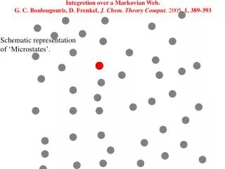
p85α SH3 p85β SH3 Figure 8 depicts a structural superposition of the SH3 domain and p85α (PDB ID: 3I5S) and the SH3 domain from p85β (PDB ID: 2KT1)using Cαatoms in pymol.