Download
1 / 71
750 likes | 1.13k Views
Biliary Tree Infection, Liver Abscess and Hepatitis A. OCTOBER 2005. Acute cholecystitis Cholangitis Liver abscess Hepatitis All cause jaundice. } Viral. Biliary Tree Infection. Bacterial Bacterial/Protozoan. Cholecystitis. Hepatitis / Liver abscess. Cholangitis. Jaundice.

E N D
Biliary Tree Infection, Liver Abscess and Hepatitis A OCTOBER 2005
Acute cholecystitis Cholangitis Liver abscess Hepatitis All cause jaundice } Viral Biliary Tree Infection Bacterial Bacterial/Protozoan
Cholecystitis Hepatitis/Liver abscess Cholangitis
Jaundice • = hyperbilirubinaemia due to various causes • Can be due to conjugated or unconjugated bilrubin • Classically presents with yellow eyes, skin, pale stools and dark urine • May have a hepatomegaly, pruritus • Can be due to obstructive (tumour, gallstones) or non obstructive causes (hepatitis, alcoholic hepatitis, haemolysis)
Bilirubin • Bilrubin is bound to albumin in blood • delivered to liver, conjugated to sugar residues to make it water soluble • secreted in bile • Once in gut bilirubin reduced to urobilinogen (by bacterial flora) • urobilinogen is required (indirectly) to give stools characteristic colour
Bilirubin If this process is blocked: • less bilirubin ends up in bile • more in blood and therefore more ends up in urine • So stools lose their pigment (pale) • and excess bilirubin in blood exits through urine giving it dark colour
Liver Enzymes • Some are a better measure of hepatocellular damage e.g. Alanine aminotransferase (ALT), Aspartate aminotransferase (AST) • This can be due to hepatitis, drugs, alcohol, toxins etc • Others are a better measure of obstruction e.g. Alkaline phosphatase, serum bilirubin • This can be due to cholecystitis, cholangitis, tumours, gall stones
Lab Test Intrahepatic(hepatocellular) Extrahepatic(obstructive) AST, ALT +++ + Alk. Phos. + +++ Bilirubin ++ +++ Cholestatic Jaundice Laboratory Evaluation
Acute Cholecystitis • Acute inflammation of gallbladder • Affects 10% of Western population • Of these only 1/5 symptomatic • Of these, 1-3% develop cholecystitis, i.e. ~ 1/2500 of general pop) • Does not always involve infection
Acute Cholecystitis Pathogenesis • Gall stones • 90% due to Obstruction of cystic duct e.g. by stones, biliary sludge, tumour or scarring • Necrosis • 20% of cases involve infection with normal bowel flora (E. coli, Klebsiella, Enterococcus spp., Bacteroides spp., Clostridiaspp.) • In developing countries, ascariasis worm major cause
Classification OEDEMA OEDEMA plus CONGESTION FOCAL NECROSIS SUPPURATION GANGRENE PERFORATION INTRAMURAL INTRALUMINAL PERI-CHOLECYSTIC LOCALISED FREE
Gall Stones • Gall stones can consist of bile salt, cholesterol or be mixed
Prostaglandins mediate inflammatory response, vicious circle
Acute Cholecystitis Clinical Features • Nausea, vomiting, fever • Constant pain in right upper quadrant of abdomen • Murphy’s sign (pain preventing full inspiration during subhepatic palpation) • +/- jaundice • +/- palpable gall bladder • Temperature • Said to occur classically in women who are “fair, fat and forty with four children” but can occur in anybody • If infection occurs, may have signs of septicaemia (worse prognosis) or peritonitis if perforation occurs
Acute Cholecystitis Diagnosis • Based on Clinical Features and Investigations • Radiological findings US, PFA, • Lab findings, increased WCC, ESR, CRP
Radiological Investigations • Ultrasound scan – can reveal stones, oedema of gall bladder wall, fluid around gall bladder • Plain film of abdomen (X-ray) shows stones in 10%, may show air level in emphesymatous cholecystitis • If US unclear, Scintigraphy used. Inject radiolabelled HIDA which is secreted in bile. Failure of appearance in gallbladder within 1-2 hours is a sign of blockage • CT may also be useful
Ultrasound Gallstones seen on X-ray
Organsims Involved • Usually mixed, derived from bowel flora • Enterobacteriaceae 68% • Enterococci 14% • Bacteroides spp. 10% • Clostridium sp. 7%
Acute Cholecystitis Complications • Perforation, necrosis gangrene, suppuration • Abscess formation • Septicaemia • Peritonitis
Acute Cholecystitis Treatment • Most respond to conservative management, gallstones falls back into gall bladder, cystic duct empties and symptoms resolve • Rest gall bladder – no food, IV fluids, pain relief as required • Indomethacin to reduce prostaglandin effects • If systemic signs or no improvement after 12-24 hours give Antibiotics, usually: • Co-Amoxiclav or Ampicillin, Ciprofloxacin or Gentamicin , and Metronidazole • Piperacillin/Tazobactam (Tazocin)
Acute Cholecystitis Treatment • About 20% require emergency surgery • These are patients with deterioration in condition,perforation with peritonitis, suspected pericholecystic abscess or emphysematous cholecystitis • Many others will require surgery but timing of surgery is a matter of debate • Cholecystectomy = removal of gallbladder, can be done as an open or laparoscopic procedure
Cholangitis Definition • continuous, varying degrees of inflammation and /or infection involving hepatic and common bile duct (mucosa continuous) More severe entities are ascending cholangitis and acute obstructive suppurative cholangitis. Pathogenesis • Essentially obstruction of the common bile duct due to stones, parasites, surgery, leading to increased pressure, oedema, congestion, necrosis and proliferation of bacteria.
Ascending Cholangitis Signs and Symptoms • Previous history of gall bladder disease, • acute onset of Charcot's triad • RUQ pain, • fever & rigors • Jaundice Treatment • Antibiotics e.g.Co-Amoxiclav or Ampicillin ,+Ciprofloxacin or Gentamicin ,+ metronidazole or Piperacillin-tazobactam+/-Ciprofloxacin • Decompression by endoscopy(ERCP), radiological stenting or surgical drainage • Delay in treatment can result in septicaemia, liver abscess
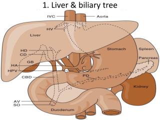
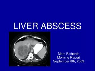
Liver Abscess • Liver is a very vascular organ, receiving blood from systemic and portal circulation • Bile drainage also provides route of bacterial entry especially when obstruction occurs • Usually Kuppfer cells lining hepatic sinusoids clear bacteria to prevent infection
Liver Abscess Other causes include trauma and secondary infection also crytogenic Liver abscess can be pyogenic or amoebic
Liver Abscess • Untreated pyogenic liver abscess is uniformly fatal • Appropriate antibiotic Tx and drainage reduces mortality to 5-30% • Abscess can be single or multiple ( due to biliary disease) • Right lobe being bigger is more commonly involved
Liver Abscess Clinical Features • Fever, chills for several days or weeks • Spiking temperatures with ascending cholangitis • Malaise • Anorexia • Weight loss • +/- referred pain to right shoulder
Liver Abscess Clinical Findings • hepatomegaly • +/- tenderness • Reduced breath sounds on right hand side • Hepatic friction rub • Jaundice in 25%
Most Common Causative Organisms Usually mixed aerobes and anaerobes, type often corresponds to source: • Gram negative bacilli from GIT e.g E.coli, Klebiella spp • Streptococcus milleri from GIT • Bacteroides, Fusobacterium and other anaerobes from GIT • S. aureus from haematogenous spread • Entamoeba histolytica • Fungal abscesses e.g. C. albicans in patients with prolonged antibiotic exposure, transplants, immunocompromised patients
Liver Abscess Investigations • FBC: anaemia, raised WBCs • Raised CRP and ESR • Raised liver enzymes esp Alk Phos • Blood cultures • Culture abscess fluid • Radiology: CT or ultrasound
Liver Abscess Treatment • Percutaneous drainage of abscess under CT or US guidance • If this fails – surgical drainage • Exception is Entamoeba histolytica which responds to metronidazole without surgery • CT/US to monitor Tx
Liver Abscess Antibiotic Treatment depends on culture results: • Ampicillin+Gentamicin + metronidazole orPiperacillin-tazobactam • Clindamycin, Flucloxacillin for sensitive Staphylococci • Amphotericin B for fungi • Entamoeba histolytica – metronidazole • Usually for 1-4 months
Hepatitis Viruses • Types A to G • No relation to each other, simply infect same organ • Viral hepatitis can also be caused by other viruses e.g. EBV, CMV and HSV
Hepatitis A - History • “Catarrhal jaundice” recognised in ancient China, Greece and Rome Hippocrates • Epidemic in Minorca in 1745 • McDonald first to suggest viral cause in 1907
Viral Hepatitis - History • Deliberate transmission to human volunteers in Germany in 1942 • Jaundice committee 1943, One(serum) has incubation period 60-160 Hep B and another feacal oral route , I.P shorter Hep A identified • Mid 1970`s- new serological test for Hepatitis B did not explain all cases-nonA-nonB now Hepatitis C (1989) but sporadic and community acquired Hep E also described • Rizzetto 1977 described Hep D while working on Hep B • Other causes of jaundice complicated early understanding and were separated into “infectious hepatitis” and “serum hepatitis”
Hepatitis A - Classification • Picornaviridae of which there are 3 genera: • Rhinovirus (Rhinoviruses) • Enteroviruses (Polio, coxsackie, echo and enteroviruses) • Hepatavirus (Hepatitis A) • RNA virus, ss + RNA (7.5 kb) • Only one serotype • Non-enveloped • 27-28 nm icosahedral structure
Hepatitis A Virus (HAV) • First isolated in 1979 • Natural host: human • Stable: heat and acid-resistant • Inactivated by high temperature, formalin, chlorine
Hepatitis A – Life Cycle • Infected material ingested • Absorbed through stomach or small intestine • Replicates in liver • Secreted into bile • Excreted into stool or reabsorbed • Spread: Faecal-oral route
Hepatitis A Robust virus: • stable after incubation at 56oC • Lasts for years at –20oC • In dried form at room temperature can last for several weeks • Killed by boiling for 5 minutes
Hepatitis A • Therefore steaming shellfish probably insufficient • Survives for days/months in live oysters, waste water, soil • Stable at pH 3 • Resistant to diethyl ether, chloroform and 50% trichlorotrifluorethane
Hepatitis A • i.e. tough organism which, because of its ability to survive, is easily transmitted • Therefore meticulous care is needed when handling clinical specimens • Destroyed by: • Boiling • Autoclaving • Chlorine • Iodine • Radiation • formaldehyde
HAV Epidemiology • Man is natural host • Worldwide distribution • Late Autumn , early Winter • Virus spread in feces • Virus contaminates • Water: drinking, bathing; washing food • Food: shellfish and other filter feeders • Hands: personal hygiene; contaminated water
Hepatitis A - Epidemiology • CDC 10,600 cases in USA/2001, 22% hospitalised, ~ 100 deaths/yr • Over last 40 years average age of infection increasing due to improved sanitation, detection and prevention • Approx 10% of children and 40% of adults will have IgG • Highest rate of seropositivity in Africa, Asia and South America • But epidemiology is changing with improved hygiene and increasing travel