Download
1 / 28
280 likes | 398 Views
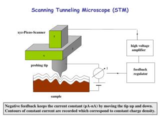
Photonics for bio-imaging and bio-sensing Rongqing Hui Dept. Electrical Engineering & Computer Science, The University of Kansas, Lawrence Kansas. Fluorescently labeled tissue. 3-demensional translation stage. Detector pin-hole. Photo detector. Laser source. Lens. Lens. Beam splitter.

E N D
Photonics for bio-imaging and bio-sensingRongqing HuiDept. Electrical Engineering & Computer Science,The University of Kansas, Lawrence Kansas
Fluorescently labeled tissue 3-demensional translation stage Detector pin-hole Photo detector Laser source Lens Lens Beam splitter Lens Focal plane Out of focus Laser scanning confocal microscope Advantages: out-of-focus background can be removed by the small aperture in front of the detectorAllows 3-D imagingDisadvantages:Photon bleach because the use of visible light (400 – 600 nm)sensitive to background light
Excited state Excited state ~800nm Fluorescence Fluorescence ~400nm ~800nm Ground state Ground state One photon excitation Two photon excitation Concept of Two-photon excitation
Optical power spreading Better focus Two-photon microscopy Use near infrared wavelength -less photon bleach and less scattering when penetrating through tissueDetection at wavelength far away from excitation – no background noise due to Raman and direct fluorescenceTwo-photon excitation is proportional to the square of the power density – smaller focus point, minimum off-focus excitation and no need of a pin-hole in front of the detector
Why two-photon microscopy is not popular so far ? • Requires very high peak optical power because of the low 2-photon excitation efficiency • Ti:Sapphire lasers have to be used to provide kW level peak optical power: big in size, very expensive, needs tweaking from time to time • Difficult to deliver femtosecond optical pulse from laser to microscope
Saturable absorption mirror Faraday rotator Doped MM fiber Faraday rotator Partial reflection Pump High power femtosecond fiber laser Popular wavelengths: 1550nm: Erbium doped fiber 1064nm: Ytterbium doped fiber 780nm: Frequency-doubling the output of 1550nm fiber laser using periodically polled LiNbO3 (PPLN)
Enabling technologies • Improvement in rear-earth doped optical fibers • Excite only the fundamental mode of a doped multi-mode fiber: breakthrough power limitations • Use Faraday rotator: eliminate polarization sensitivity • Saturable absorption mirror: pulse shaping
Highly nonlinear Photonic crystal fiber • Periodic air holes in the core • Very high nonlinearity • Zero-dispersion wavelength shifted to 700nm • Support Raman shifted soliton in NIR
6m photonic crystal fiber Optical spectrum analyzer Femtosecond fiber laser Mechanical translation stage Wavelength shift using photonic crystal fiber
Power spectral density (linear) Wavelength (nm) Pulse wavelength shift due to power change(from 1mW to 4mW average power) Fundamental soliton condition:
Pulse wavelength shift due to power change(Computer simulation)
Microscope Source PCF-1 Filter 780 nm fiber laser AOM Pulse compressor Sample PCF-2 1050 nm fiber laser AOM Detector Digital control Register Image @ l1 Memory & signal processing Image @ l2 Control and signal processing Image @ ln Fig.3. Block diagram of the proposed wavelength switchable two-photon equipment Multi-color two-photon fluorescent microscopy using TDM
At depth 2 At depth 1 Measured Two-photon fluosphere images
Radial and axial two-photon intensity profiles(excited at 780nm)
Radial and axial two-photon intensity profiles(excited at 920nm)
Observation volume area Focal volume area Fluorescent correlation spectroscopy (FCS)(measured at 780nm)
Bio-assay based on flow cytometryCount and analyze individual particles in a fluid channel
UV Photo-mask Spin-coat SU-8 photo-resistor Silicon substrate Silicon substrate Flow-cytometer on chip:Create channels
UV Photo-mask Spin-coat second layer SU-8 Silicon substrate Flow-cytometer on chip: Create grooves Silicon substrate
PDMS Molding PDMS Silicon substrate
Input fiber Detection fiber Buffer Buffer Detection fiber Sample Picture of a flow chip
Buffer Buffer Sample Demonstration of sheath flow Sheath creation
Computer D/A converter Laser PMT Fluid pump (water) Fluid pump (water) Waste Fluid pump (sample) Measurement
PMT output (V) Time (s) Measurementpassing diluted yeast solution through the cytometer
Clean water 4 times increase of yeast concentration between each measure
10log(N) 10log(V) Cumulative summation histogram V: threshold n: number of counts