Download
1 / 39
420 likes | 451 Views
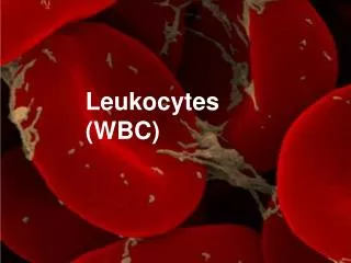
Lecture topics: characteristics and functions of WBCs*. Types of leukocytes Production ( Leukopoiesis ) Functional characteristics of the different leukocytes Mechanisms: How do they do what they do?. Leukocytes.

E N D
Lecture topics: characteristics and functions of WBCs* • Types of leukocytes • Production (Leukopoiesis ) • Functional characteristics of the different leukocytes • Mechanisms: How do they do what they do?
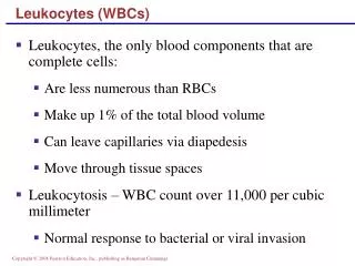
Leukocytes • Only formed elements that are complete cells with a nucleus and cytoplasmic organelles. • On average, there are 4000-10000 WBC/l of blood. Average no.=7000 l of blood • Remember they account for about 1% of total blood cells. • Leukocytosis: An increase in no. of WBCs in circulation. - specific WBC type: lymphocytosis, granulocytosis, neutrophilia. • Leukopenia: A decrease in no. of circulating WBCs.
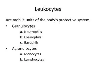
Types of leukocytes** • Neutrophils………62% • Eosinophils………2.3% • Basophils…………0.4% • Monocytes………..5.3% • Lymphocytes……..30% Polymorphonuclear Or granulocytes agranulocytes Note: To remember the order of abundance leukocytes from most abundant to least abundant: Never Let Monkeys Eat Bananas **granulocytes and monocytes protect body by ingestion (phogocytosis) of invading organisms( for example bacteria). They are formed in bone marrow. ** lymphocytes protects body through immune system. They are formed in lymphogenous tissues (lymph glands, spleen, tonsils, thymus, Peyer’s patches in intestinal wall.
Genesis of WBCs (leukopoiesis) Pluripotential stem cell myeloblast lymphoblast T lympho B lympho Plasma cell neutro mono baso eosino
Life span of WBCs* • Granulocytes stay in blood for 4-8 hours and in the tissues 5 days. When there is serious infection their whole life span is shortened to few hours • Monocytes stay in the blood for 10-20 hours and then they leave to different tissues where they form tissue macrophages. Tissue macrophages can live for months. If there is infection these macrophages may die earlier. • Lymphocytes circulate between blood and lymphoid tissues through lymph and they survive for weeks and months
Neutrophils and macrophages defend against infectionMacrophage : Monocytes in the blood then in the tissues swell and increase the diameter and become
Leukocytes cross capillary wall by Diapedesis Diapedesis • Leukocytes able to slip out of the capillary. They sequeeze themselves through small pore between endothelial cells that form the capillary wall. • All leucocytes can make this movement between blood and interstitial space. Note: diapedesis is increased in inflammation. Factors released from inflamed tissues make WBCs to stick to the capillary wall in inflamed tissue (this effect is called margination). And this increases diapedesis.
Amoeboid motion of Leukocytes • leukocytes move through the tissue spaces through this motion. • They form cytoplasmic extensions that move them along (pseudopods). These pseudopods also help WBC to phogocytize foreign particles.
Movement of WBCs by Chemotaxis towards an area of tissue damage • Chemical substances (chemotactic substance, chemotaxins) found and produced in the tissues or released by other leukocytes that attract WBCs toward these chemicals. • Chemotaxins include 1. Bacterial or viral toxins • Degenerative products of inflamed tissues • Reaction products of ‘complement complex’ • Reaction products of plasma clotting Chemotactic signals effective till 100m
Opsonization • Plasma factors act on the bacteria to make them ‘tasty’ to the Phagocytes. It enhances the phagocytic ability of WBCs • Opsonins which coat the bacteria are IgG and Complement proteins • Coated bacteria bind to receptors on the Neutrophil membrane. bacteria
Phagocytosis • Means ingestion of invading agents (bacteria, viruses, foreign bodies, dead tissues). Done mostly by macrophages and neutrophils (neutrophils can phagocytize 3-20 bacteria and macrophages can phagocytize up to 100 bacteria) • 1. Selective – Natural structures is protected from phagocytosis by smooth surface and protective proteins. While foreign bodies have rough surfaces and no protective proteins so they are liable to be phagocytized. • 2. Antibodies adhere to surface of bacteria and make them susceptible for phagocytosis (opsonization).
Phagocytosis by neutrophils and macrophages • Phagocytized particles digested by intracellular enzymes • Neutrophils and Macrophages have proteolytic enzymes. • Macrophages (but not neutrophils) have lipases which can digest lipid membrane surrounding some bacteria (Tuberculosis bacilli) • Macrophages are more powerful phagocytes than neutrophils. They can engulf large particles and digest them and extrude the residual products of digestion so they survive after so many phogocytosis process.
Phagocytosis process • The WBC sends pseudopods around foreign particle and making a enclosed chamber that contains the particle forming phagocytic vesicle (called phagosome). • Lysosomes containing digestive enzymes fuse with phagosome which is called now digestive vesicle. The particle is digested and residual products are thrown outside WBC.
Phagocytosis (cont.) • Neutrophils and Macrophages produce Oxidizing agents which are bactericidal (killing bacteria) e.g. Super oxide(O2-), Hydrogen peroxide(H2O2), hydroxyl ions • Myeloperoxidase is lysosomal enzyme which can convert chloride to form hypochlorite which is very strong bactericidal agent. Note: O2- and H2O2 are Oxidants and Bactericidal
Reticuloendothelial system RES: Monocytes, Mobile macrophage, fixed macrophage, spleen, lymph nodes and specialized endothelial cells in the bone marrow (all originated from monocytic stem cells)RES is used in medical terms than monocyte macrophages system as they are synonymous
Special macrophages are present in :1. Skin, subcutaneous tissues2. Lymph nodes3. Alveolar macrophages in the lungs4. Macrophages (Kupffer cells) in the liver 5. Macrophages in the spleen and bone marrow
Eosinophils • About 2% of all leukocytes • Ineffective protection against usual types of infection. • Contain large granules that stain red. • About the same size as neutrophils but weaker phagocytes • nucleus has 2 lobes
Eosinophils (cont.) • Eosinophils no. is increased in people with parasite infection (like worms) • Release the enzymes (hydrolytic enzymes, highly reactive form of oxygen, major basic protein) from their cytoplasmic granules onto the parasite’s surface, killing the parasites . • Eosinophils are attracted by chemotactic factor released by basophils and mast cells toward allergic tissues. • They detoxify the substances that released during allergic reaction. Also they engulf antibodies-antigens complexes so they help in reducing spread of allergic reaction
Basophils • Large histamine-containing granules • Affinity for basic dyes so they stain purplish black (baso = basic). • Similar in function to mast cells. Mast cells are found in the tissues. • Both mast cells and basophils liberate histamin and heparin, small quantities of bradykinins and serotonin.
Basophils antigen Basophils and mast cells play important role in allergic reactions. IgE attached to surface of basophils and when antigen bind with IgE on the surface, degranulation follows this binding and release of histamine, bradykinin, serotonin, Slow Reactive Substance of Anaphylaxis, heparin and other lysosomal enzymes
Agranulocytes monocyte • Lack visible cytoplasmic granules • Include lymphocytes and monocytes B lymphoctes • Attack bacteria, viruses and toxins and converted into a. Plasma cells that produce antibodies (Ig = immunoglobulin) b. Memory cells • Keep a “record” of pathogens
T Lymphocytes • T cells • Acting directly against virus-infected cells and tumor cells 1. CD 4 • T helper cells 2. CD 8 • T cytotoxic cells 3. suppressor T cells
1. T Helper cells : (AIDS) The most numerous type of T cells. They help in the function of immune system , forming protein mediators called lymphokinasewhich cause:a. Stimulation of growth and proliferation of cytotoxic and suppressor T cellsb. Stimulation of B cells growth and form plasma cells and antibodies.
c. Activation of macrophage system.d. Feedback stimulatory effect on the T helper cells (positive feedback effect
2. Cytotoxic cells : Killer cells, direct attack3. Suppressor cells: Limiting the ability of immune system to attack a person’s own body tissues (Immune tolerance)
Monocytes • They have abundant pale-blue cytoplasm and a darkly staining purple nucleus, which is distinctively U or kidney shaped • Migrate from blood into tissues where they enlarge and become macrophages (which are phagocytic). • Most powerful phagocyte • They can phagocytize whole RBC, malarial parasites and dead neutrophils • They are very important in starting immune responses • They can be called different names when they stay in different tissues. • Skin…..histiocytes • Lungs...alveolar macrophage • Liver….Kupffer cells
Plasma cellA type of immune cell that makes large amounts of a specific antibody. They are develop in the bone marrow from B cells that have been activated.They are transported by plasma and lymphatic system
Mast cell:Originate from bone marrow (Monocyte or lymphocytes??),very similar to basophil.Found widely in the body particularly in connective tissues. They produce histamine, Heparin, serotonin,Dopamine and also involve in the immunological, allergic and inflammatory conditions.
Leukocytosis: An increase in no. of WBCs in circulation. - specific WBC type: lymphocytosis, granulocytosis, neutrophilia. Leukopenia: A decrease in no. of circulating WBCs. Irradiation, Xray or gamma rays. Drugs, chloramphenicol, thiouracil , borbiturate hypnotics
Leukemia :Uncontrolled production of WBC 1.Lymphocytic (Lymph nodes)2. Myelogenous (Bone marrow)
Effects of leukemia:Infection, Sever anemia, Bleeding tendency, Excessive use of metabolic substrate by cancerous cells cause deblitate of other tissues.
Role of macrophage and neutrophils in inflammation • When bacteria get access to certain tissue, the first line of defense is macrophages which are already in that tissue. • Within hour or so, the no. of neutrophils starts to increase in the area. Neutrophils form the second line of defense.. These neutrophils come from blood due to liberation of chemotactic substances.
Role of macrophage and neutrophils in inflammation (cont.) • In severe infection the no. of neutrophils (neutrophilia) in the blood increases dramatically 3rd. Products of inflammation stimulate bone marrow to release the stored neutrophils there in bone marrow 4th. ( remember that bone marrow stores of WBCs is more that no. of WBCs in the blood).
Role of macrophage and neutrophils in inflammation (cont.) • Monocytes from blood enter the site of inflammation and become active macrophages in few days. • Factors (Substances) released from activated macrophages stimulate bone marrow to produce new monocytes and neutrophils.
Role of macrophage and neutrophils in inflammation (cont.) • Monocytes from blood enter the site of inflammation and become active macrophages in few days. • Factors (Substances) released from activated macrophages stimulate bone marrow to produce new monocytes and neutrophils. Activated macrophage Releasing 1.TNF (tumor necrosis factor 2. GM-CSF (granulocyte monocyte colony stimulating factor 3. M-CSF (monocyte colony stimulating factor 4. G-CSF (granulocyte colony stimulating factor Granulocytes, monocytes
Role of macrophages in activation of lymphocytes • When macrophage phagocytize invading microorganisms and digest them, the antigenic products liberated into macrophage cytoplasm. Macrophages pass these antigens directly to lymphocytes leading to activation of specific lymphocytes that can make proper immune reaction against invading microorganism. • Also macrophages produce substance (interleukin-1) that stimulates growth and reproduction of specific lymphocytes. bacteria macrophage lymphocyte
Character of inflammation • Vasodilatation, permeability, interst fluid clotting, increase no of granulocyte and monocyte, swelling of the tissue. • Tissue product: Histamine, bradykinin, serotonin, prostaglandin. • (Walling off) effect of inflamation.
Feedback control of neutro and macro TNF, Interleukin 1(IL-1), GM-CSF, M-CSF, G-CSF These factors are formed by activated macrophages in the inflamed tissues
Formation of pusNeut and Macro engulf bacteria and necrotic tissues then Neut and macro die and cavity formed in the inflamed tissue. This cavity contains necrotic tissue, dead Neut, dead Macro and tissue fluid. This mixture known as pus