Download
1 / 129
1.29k likes | 1.32k Views
PULMONARY INFECTIONS. Prof.Dr.Ferda ÖZKAN. Normal Lung. The lungs are constructed to carry out their cardinal function: the exchange of gases between inspired air and blood. Normal Lung. The respiratory system is an outgrowth from the ventral wall of the foregut.

E N D
PULMONARY INFECTIONS Prof.Dr.Ferda ÖZKAN
Normal Lung The lungs are constructed to carry out their cardinal function: the exchange of gases between inspired air and blood.
Normal Lung • The respiratory system is an outgrowth from the ventral wall of the foregut. • The midline trachea develops two lateral outpocketings, the lung buds. • The right lung bud eventually divides into three branches-the main bronchi-and the left into two main bronchi, thus giving rise to three lobes on the right and two on the left. • The lingula on the left is the middle lobe equivalent; however, the left lung is smaller than the right.
Normal Lung • The right main stem bronchus is more vertical and more directly in line with the trachea than is the left. Consequently, aspirated foreign material, such as vomitus, blood, and foreign bodies, tends to enter the right lung rather than the left.
Normal Lung • The main right and left bronchi branch give rise to progressively smaller airways. • Progressive branching of the bronchi forms bronchioles, which are distinguished from bronchi by the lack of cartilage and submucosal glands within their walls.
Normal Lung • Further branching of bronchioles leads to the terminal bronchioles, which are less than 2 mm in diameter. The part of the lung distal to the terminal bronchiole is called the acinus; it is approximately spherical, with a diameter of about 7 mm.
Normal Lung • An acinus is composed of respiratory bronchioles , which give off several alveoli from their sides. • These bronchioles then proceed into the alveolar ducts, which immediately branch into alveolar sacs, the blind ends of the respiratory passages, whose walls are formed entirely of alveoli, which are the site of gas exchange. • The alveoli open into the ducts through large mouths. All alveoli are open and have incomplete walls. A cluster of three to five terminal bronchioles, each with its appended acinus, is usually referred to as the pulmonary lobule.
Normal Lung • The entire respiratory tree, including the larynx, trachea, and bronchioles, is lined by pseudostratified, tall, columnar, ciliated epithelial cells, heavily admixed in the cartilaginous airways with mucus-secreting goblet cells- except for the vocal cords, which are covered by stratified squamous epithelium,
The microscopic structure of the alveolar walls • The capillary endothelium lining the intertwining network of anastomosing capillaries. • A basement membrane and surrounding interstitial tissue separating the endothelial cells from the alveolar lining epithelial cells. In thin portions of the alveolar septum, the basement membranes of epithelium and endothelium are fused, whereas in thicker portions, they are separated by an interstitial space (pulmonary interstitium) .
Alveolar epithelium, which contains a continuous layer of two principal cell types: • flattened, platelike type I pneumocytes (or membranous pneumocytes) • covering 95% of the alveolar surface and rounded type II pneumocytes. Type II cells are important for at least two reasons: (1) They are the source of pulmonary surfactant, contained in osmiophilic lamellar bodies seen with electron microscopy, and (2) they are the main cell type involved in the repair of alveolar epithelium after destruction of type I cells.
Alveolar macrophages, loosely attached to the epithelial cells or lying free within the alveolar spaces, derived from blood monocytes and belonging to the mononuclear phagocyte system. Often, they are filled with carbon particles and other phagocytosed materials. • The alveolar walls are not solid but are perforated by numerous pores of Kohn, which permit the passage of bacteria and exudate between adjacent alveoli
Introduction • Daily 10,000 liters of air – filtered • Nasopharyngeal flora (during sleep) • Virulent organisms. • Pneumonia: Inflammation of lung. • Respiratory tract infections – commonest in medical practice. • Enormous morbidity & mortality.
Decreased resistance - General/immune Virulent infection - Lobar pneumonia Defense MechanismsIn the normal respiratory system there are a number of important defense mechanisms that protect the lung from infection. These include: Reflex closure of the vocal cords Cough reflex Mucociliary clearance Macrophage activity and immune competence. Etiology
An increased risk of bacterial infection is associated with impairment of the defense mechanism, as in any of these clinical situations: • Loss of consciousness • Immunodeficiency state • Pulmonary edema • Neutropenia • Chronic airway obstruction • Viral infection.
ExudateThe exudate in bacterial pneumonia is typically composed of varying proportions of: • edema fluid • red blood cells • leukocytes (principally neutrophils) • fibrin • The cellular exudate in acute bacterial pneumonia is in the alveolar spaces and distal bronchioles though in severe cases the major airways may also be filled with purulent secretion.
Viral Bacterial Mycoplasmal Fungal Types
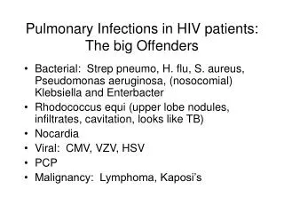
Community-acquired acute pneumonia Streptococcus pneumonia Haemophilus influenza Moraxella catarrhalis Staphylococcus aureus Legionella pneumophilia Klebsiella Pseudomonas Community-acquired atypical pneumonia Mycoplasma Chlamydia Legionella Viruses (RSV, parainfluenza & influenza, adenovirus) Nosocomial pneumonia Gram negative rods Staphlyococcus aureus Aspiration pneumonia Anaerobic oral flora Amniotic fluid Gastric content Chemicals Chronic pneumonia Nocardia Actinomyces Granulomatous Necrotizing pneumonia Anaerobic Staphlyococcus aureus Klebsiella Streptococcus pyogens The Pneumonia Syndromes
Patterns of Pulmonary infections • Airway – Bronchitis/Bronchiolitis, Bronchiectasis • Parenchyma • Pneumonia • Bronchopneumonia • Lobar pneumonia • Lung abscess • Tuberculosis
Routes of Infection Several possible routes of infection of the lung exist: • Aspiration of contaminated secretions--most common • Inhalationof infected airborne droplets • Bacteremia • Direct extension of an acute inflammatory process from an adjacent organ or structure.
Etiopathogenesis • Causes of bacterial pneumonia can be categorized as extrinsic and intrinsic. • Extrinsic factors: infection with respiratory pathogens. Exposure to pulmonary irritants or direct pulmonary injury causes noninfectious pneumonitis. • Infectious agents responsible for bacterial pneumonias include S. pneumoniae and H. influenzae;Klebsiella, Staphylococcus, and Legionella species; and gram-negative organisms. • Aspiration and inhalation of aerosols containing the bacterial pathogen are the most common modes of infection. • Some bacteria, such as Staphylococcus species, may spread to the lungs hematogenously.
S. pneumoniae is the most common cause of bacterial pneumonia. • Pneumonia from H influenzae often is associated with debilitating conditions such as asthma, COPD, smoking, and a compromised immune system. • K. pneumoniae may cause a severe necrotizing lobar pneumonia in patients with chronic alcoholism, diabetes, or COPD. • S. aureus pneumonia is observed in those who abuse intravenous drugs. • S. aureus generally occurs in hospitalized patients and patients with prosthetic devices; it spreads hematogenously to the lungs from contaminated local sites. This pathogen also is an important cause of pneumonia following infection with influenza A. • L. pneumophila infections occur either sporadically or as local outbreaks.
Gram-negative pneumonias are observed in individuals who are immunocompromised or hospitalized. • Causative organisms include Escherichia coli and Pseudomonas, Enterobacter, and Serratia species. Residents of chronic care facilities are at risk for gram-negative pneumonia.
Intrinsic factors: related to the host's immune response, the presence of comorbidities, and other risk factors: • Loss of protective reflexes allows aspiration of oropharyngeal flora into the lung. • Aspiration is facilitated by altered mental status from intoxication, deranged metabolic states, neurological causes (eg, stroke), and endotracheal intubation. • Local lung pathologies (eg, tumors, chronic obstructive pulmonary disease [COPD], bronchiectasis). • Smoking impairs the host's defense to infection by a variety of mechanisms.
Aspiration pneumonia is observed in individuals with altered sensorium (eg, seizures, alcohol intoxication, drug intoxication) or CNS impairment (eg, stroke). • The stomach or oropharyngeal contents are aspirated.
Complications of Pneumonia • Destruction of lung tissue from infection (leading to bronchiectasis) • Organization of the exudate • Abscess formation • Spread of the infection to the pleural cavity (empyema) • Sepsis & Pyemia • Respiratory failure • Acute respiratory distress syndrome • Superinfection with gram-negative organisms • Death
1. Pneumonia • 1.1. Bronchopneumonia • 1.2. Lobar pneumonia • 1.3. Viral (Atypical) pneumonia
1.1. Bronchopneumonia • Staph, Strep, Pneumo & H. influenza • Bronchopneumonia is characterized by focal areas of suppurative inflammation, in a patchy distribution, involving one or multiple lobes. • Usually bilateral • Lower lobes common • Complications: • Abscess • Empyema • Dissemination.
The inflammatory exudate in each of the foci of involvement typically involves asmall airway and surrounding alveolar spaces. • Histologically • The relatively small areas of involvement • Focal areas of suppurative inflammation in a patchy distribution. • Abscess formation.
Bronchopneumonia Patchy consolidation – not limited to lobes
1.2. Lobar Pneumonia • Fibrinosuppurative consolidation – whole lobe • Rare (due to antibiotic treatment) • ~95% - Strep. pneumoniae types 1,3,7& 2 • Four stages (Laennec,1838) : • Congestion & edema (1 to 2 days) • Red Hepatization (2-4 days ) • Gray Hepatization (4 to 8 days) • Resolution (1 to 3 weeks).
Congestion & Edema: This stage is characterized histologically by: • vascular engorgement, • intra-alveolar fluid, • small numbers of neutrophils, • often numerous bacteria. • Grossly, the lung is heavy and hyperemic.
Red hepatization: • Vascular congestion persists, • Extravasation of red cells into alveolar spaces, • Increased numbers of neutrophils and fibrin. • The filling of airspaces by the exudate leads to a gross appearance of solidification, or consolidation, of the alveolar parenchyma. • A dry, granular, dark-red lung surface on gross appearance • This appearance has been likened to that of the liver, hence the term "hepatization".
Gray hepatization: • As pneumonia progresses over 2-3 days, erythrocytes are lysed with persistence of the neutrophils and fibrin and, epithelial cells degenerate • The alveoli still appear consolidated, but grossly the color is paler and the cut surface is drier.
Resolution: • The exudate is digested by enzymatic activity, and cleared by macrophages or by cough mechanism. • Dying pneumococci release a preformed toxin, further contributing to this damage. • The pneumococci are opsonized by leukocytes and begin to be cleared. • Resolution results in the formation of jellylike yellowish-colored exudates. • Absorption of these exudates is remarkably efficient, with little organization or permanent scaring.
Strep. Pneumoniae Pneumonia • Streptococcus pneumoniae produces few toxins. • It causes diseases by its capacity to replicate in host tissues. • The presence of a capsule allows an escape from phagocytosis, resulting in an intense inflammatory response in hosts who are immunologically naive. • Colonization of the oropharynx by bacterial adherence to human pharyngeal cells is the usual first step.
The alternative pathway of the complement is first activated. • Anticapsular antibodies are effective in providing protection against pneumococcal infection. • They appear 5-8 days after the onset of infection. By this time, fever usually disappears in the absence of treatment. • Natural immunity follows infections as well as colonization.
Conditions that predispose the patient to pneumococcal infection • Defective antibody formation • Agammaglobulinemia and hypogammaglobulinemia, multiple myeloma, chronic lymphocytic leukemia, lymphoma, and HIV infection • Defective complement (C1 to C4) • Defective splenic function • Asplenia and splenectomy • Chronic diseases • Chronic obstructive pulmonary disease, cirrhosis of the liver, and alcoholism • Acute viral respiratory infections • Post–influenza virus
Pneumonia: • The absence of predisposing factors is rare in pneumococcal pneumonia affecting elderly children, teenagers, and adults younger than 60 years. • Meningitis: • Predisposing factors include head trauma, cerebrospinal fluid leak, and respiratory tract infection.