Download
1 / 24
260 likes | 635 Views
Pathology of Pulmonary Infections. Prof. Frank Carey. Topics. Pneumonia Chronic infection – abscess/bronchiectasis Tuberculosis The immunocompromised host. Pneumonia. Gr. “disease of the lungs”

E N D
Pathology of Pulmonary Infections Prof. Frank Carey
Topics.. • Pneumonia • Chronic infection – abscess/bronchiectasis • Tuberculosis • The immunocompromised host
Pneumonia • Gr. “disease of the lungs” • Infection involving the distal airspaces usually with inflammatory exudation (“localised oedema”). • Fluid filled spaces lead to consolidation
Classification of Pneumonia • By clinical setting (e.g. community acquired pneumonia) • By organism (mycoplasma, pneumococcal etc) • By morphology (lobar pneumonia, bronchopneumonia)
Organisms • Viruses – influenza, parainfluenza, measles, varicella-zoster, respiratory syncytial virus (RSV). Common, often self limiting but can be complicated • Bacteria • Chlamydia, mycoplasma • Fungi
Lobar Pneumonia • Confluent consolidation involving a complete lung lobe • Most often due to Streptococcus pneumoniae (pneumococcus) • Can be seen with other organisms (Klebsiella, Legionella)
Clinical Setting • Usually community acquired • Classically in otherwise healthy young adults
Pathology • A classical acute inflammatory response • Exudation of fibrin-rich fluid • Neutrophil infiltration • Macrophage infiltration • Resolution • Immune system plays a part antibodies lead to opsonisation, phagocytosis of bacteria
Complications • Organisation (fibrous scarring) • Abscess • Bronchiectasis • Empyema
Bronchopneumonia • Infection starting in airways and spreading to adjacent alveolar lung • Most often seen in the context of pre-existing disease
Clinical Context • COPD • Cardiac failure (elderly) • Complication of viral infection (influenza) • Aspiration of gastric contents
Organisms • More varied – Strep. Pneumoniae, Haemophilus influenza, Staphylococcus, anaerobes, coliforms • Clinical context may help. Staph/anaerobes/coliforms seen in aspiration
Complications • Organisation • Abscess • Bronchiectasis • Empyema
Lung Abscess • Localised collection of pus • Tumour-like • Chronic malaise and fever • Context - aspiration
Bronchiectasis • Abnormal fixed dilatation of the bronchi • Usually due to fibrous scarring following infection (pneumonia, tuberculosis, cystic fibrosis) • Also seen with chronic obstruction (tumour) • Dilated airways accumulate purulent secretions
Tuberculosis • Mycobacterial infection • Chronic infection described in many body sites – lung, gut, kidneys, lymph nodes, skin…. • Pathology characterised by delayed (type IV) hypersensitivity (granulomas with necrosis)
Organisms • M. tuberculosis/M.bovis main pathogens in man • Others cause atypical infection especially in immunocompromised host. Pathogenicity due to ability; • to avoid phagocytosis • to stimulate a host T-cell response
Immunity and Hypersensitivity • T-cell response to organism enhances macrophage ability to kill mycobacteria • this ability constitutes immunity • T-cell response causes granulomatous inflammation, tissue necrosis and scarring • this is hypersensitivity (type IV) • Commonly both processes occur together
Pathology of Tuberculosis (1) • Primary TB (1st exposure) • inhaled organism phagocytosed and carried to hilar lymph nodes. Immune activation (few weeks) leads to a granulomatous response in nodes (and also in lung) usually with killing of organism. • in a few cases infection is overwhelming and spreads
Pathology of Tuberculosis (2) • Secondary TB • reinfection or reactivation of disease in a person with some immunity • disease tends initially to remain localised, often in apices of lung. • can progress to spread by airways and/or bloodstream
Tissue changes in TB • Primary • Small focus (Ghon focus) in periphery of mid zone of lung • Large hilar nodes (granulomatous) • Secondary • Fibrosing and cavitating apical lesion (cancer an important differential diagnosis
Why does disease reactivate? • Decreased T-cell function • age • coincident disease (HIV) • immunosuppressive therapy (steroids, cancer chemotherapy) • Reinfection at high dose or with more virulent organism
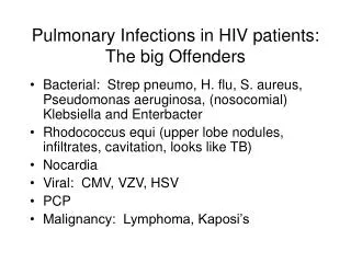
The immunocompromised host • Virulent infection with common organism (e.g. TB) • Infection with opportunistic pathogen • virus (cytomegalovirus - CMV) • bacteria (Mycobacterium avium intracellulare) • fungi (aspergillus, candida, pneumocystis) • protozoa (cryptosporidia, toxoplasma)
Diagnosis • High index of suspicion • Teamwork (physician, microbiologist, pathologist) • Broncho-alveolar lavage • Biopsy (with lots of special stains!)