Download
1 / 18
190 likes | 383 Views
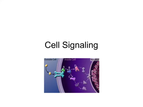
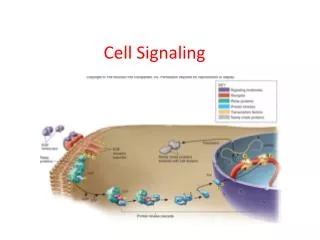
Cell Signaling: II. Protein Hardware. 1. 1. Introduction:. Numerous cytoplasmic proteins are operating in signal transduction: protein and lipid kinases and phosphatases, GTPases, and adaptor proteins .

E N D
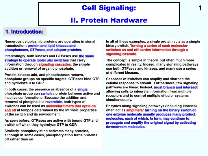
Cell Signaling: II. Protein Hardware 1 1. Introduction: Numerous cytoplasmic proteins are operating in signal transduction: protein and lipid kinases and phosphatases, GTPases, and adaptor proteins. Interestingly, both kinases and GTPases use the same strategy to operate molecular switches that carry information through signaling cascades: the simple addition or removal of organic phosphate. Protein kinases add, and phosphatases remove, phosphate groups on specific targets. GTPases bind GTP and hydrolyze it to GDP. In both cases, the presence or absence of a single phosphate group can switch a protein between active and inactive conformations. Because the addition and removal of phosphate is reversible, both types of switches can be used as molecular timers that cycle on and off at tempos determined by the intrinsic properties of the switch and its environment. As seen before, GTPases are active with bound GTP and switch off when they hydrolyze GTP to GDP. Similarly, phosphorylation activates many proteins, although in some cases, phosphorylation turns proteins off rather than on. In all of these examples, a single protein acts as a simple binary switch. Turning a series of such molecular switches on and off carries information through a signaling cascade. The concept is simple in theory, but often much more complicated in reality. Indeed, many signaling pathways use both GTPases and kinases, and many use a series of different kinases. Cascades of switches can amplify and sharpen the cellular response to stimuli. Furthermore, few signaling pathways are linear. Instead, most branch and intersect, allowing cells to integrate information from multiple receptors and to control multiple effector systems simultaneously. Enzymes along signaling pathways (including kinases) often act as amplifiers: turning on the binary switch of one enzyme molecule usually produces many product molecules, each of which, in turn, may continue to propagate and amplify the original signal by activating downstream molecules.
2 2. Protein phosphorylation: Protein kinases catalyze the transfer of the g-phosphate from ATP to amino acid side chains of proteins. Many vertebrate protein kinases phosporylate either serine or threonine. Other phosphorylate tyrosine. In eukaryotes, more than 99% of cellular phophorylation occurs on serine and threonine residues, but phosphorylation of tyrosine residues is essential for regulating many cellular processes. A few phosphorylate serine, threonine and tyrosine. Protein phosphorylation controls metabolic enzymes, cell motility, membrane channels, assembly of the nucleus, and cell cycle progression. Sometimes, it turns a process on, sometimes off. In either case, both the addition of a phosphate by a protein kinase, and its removal by a protein phosphatase are required to achieve regulation. 3. Lipid kinases: Lipid kinases phosphorylate either inositol phospholipids or a few proteins, several of which are involved with cell cycle control.
4. Protein kinases: Structure-function relationship and sub-cellular targeting: 3 The catalytic domain of protein kinases consists of about 260 residues in two lobes surrounding the ATP-binding pocket. Despite extensive sequence divergence, all of these enzymes have a similar polypeptide fold with conserved residues at critical positions required for catalysis. Each kinase has a restricted range of protein substrates, so that activation of a particular protein kinase changes the phosphorylation of a discrete subsets of proteins. Substrate specificity is achieved by selective binding of substrates to a groove that positions the acceptor amino acid at the active site, as well assecondary sites. Typically, all substrates that bind a particular kinase have similar residues surrounding the target serine, threonine, or tyrosine, (consensus sequences), allowing each substrate to interact with the same complementary residues on the kinase. Targeting of kinases to particular parts of cells also limits the range of proteins phosphorylated.
5. Effects of phosphorylation on protein structure and function: 4 Phosphorylation can inhibit protein assembly reactions. Phosphate groups can induce conformational changes or alter interactions with substrates or other molecules in several ways. • Steric interference: phosphorylation can alter the affinity of a protein for one or more of its ligands. • Example: Phosphorylation blocks substrate binding to isocitrate dehydogenase. • Surface representation with isocitrate bound to the active site. • Phosphorylation of serine 113 blocks isocitrate binding.
6. Regulation of protein kinases: 5 a. Conformational change of an activation loop: phosphorylation activates tyrosine kinases by inducing a dramatic change in a polypeptide loop that blocks the substrate binding site of the inactive enzyme. It turns on many kinases, not only tyrosine kinases. Example: Insulin receptor tyrosine kinase domain. (C): Ribbon diagram and space-filling model with the catalytic loop in orange and the activation loop in green. (D): Space-filling model of the domain, triphosphorylated on the activation loop. This rearranges the activation loop, allowing substrates (pink with a white tyrosine side chain) access to the active site. AMP-PNP is a nonhydrolyzable analogue of ATP with nitrogen bridging the b- and g-phosphates.
6. Regulation of protein kinases: 6 b. Creation of binding sites: Reversible phosphorylation controls interactions between partner proteins orintramolecular interactions which require a phosphorylated residue to complete a binding site. Example: The Src homology domain SH2 (and SH3). The Src-homoly domains (SH2 and SH3) were first recognized in Src, the first oncogene to be characterized. NT and CT lobes on the ribbon model are the N- and C-terminal lobes of the kinase domains. When tyrosine-527 is phosphorylated, the SH2 domain binds intramolecularly to the C-terminus, locking the kinase in an inactive conformation. The N-terminal SH3-domain binds intramolecularly to a proline-rich sequence (PPII-helix)connecting the SH2 and kinase domains.
6. Regulation of protein kinases: 7 c. Additional peptide sequences flanking the catalytic domain of kinases and serving special functions: • Pseudosubstrates: • These are parts of the kinase itself, or aseparate subunit, which inhibit kinases by binding to the substrate groove. • Adaptor domains: • SH2, SH3, and pleckstrin homology (PH) domains target kinases to specific sites in the cell. • Transmembrane (TM) segments: • These target receptor kinases to membranes.
6. Regulation of protein kinases: 8 d. Inhibitory phosphorylation: • Phosphorylation of myosin light chain kinase (MLCK) by protein kinase A (PKA) reduces its affinity for its protein substrate. • Phosphorylation of platelet-derived growth factor receptor kinase (by protein kinase C), inhibits its activity. e. Regulation of substrate binding: • 1. Autoinhibition: • Pseudosubstrate sequences (PSs) built into myoson light chain kinase, calmodulin activated kinase (CaMK), PKC, and protein kinase G (PKG) bind to and block the substrate sites. These autoinhibitory peptides mimic the protein substrate but lack a phosphorylatable residue. • Ca2+-calmodulin activates MLCK and CamK II by binding to a helical segment of their inhibitory peptides immediately adjacent to the pseudosubstrate site. This displaces the inhibitory peptide from the kinase and allows substrates to bind. • cGMP binding to PKG displaces the autoinhibitory peptide from the catalytic domain, activating the enzyme.
6. Regulation of protein kinases: 9 e. Regulation of substrate binding: • 2. Extrinsic regulation by inhibitory subunits: • Separateregulatory (R) subunits inhibit PKA by blocking the protein substrate site. • cAMP regulates the affinity of the regulatory subunits for the catalytic subunit. In resting cells, the regulatory subunit is free of cAMP and binds the catalytic subunit with high affinity. With a rise in cAMP concentration, cAMP binds the regulatory subunit, dissociates it from the catalytic subunit, and allows substrates access to the active site. • 3. Extrinsic regulation by activating subunits: • Regulatory subujits can also activate protein kinases. • Regulatory subun its called cyclins bind and contribute to activating cyclin-dependent cell cycle kinases, Cdks. • 4. Dual or triple regulation: • Multiple factors regulate most kinases. • Both interaction of Ca2+-calmodulin with an intrinsic pseudosubstrate and activation loop phosphorylation activate CaMK. • Both inhibitory and activating phosphorylation, as well as cyclins, regulate cyclin-dependent kinases.
6. Regulation of protein kinases: 10 f. Targeting: • Several mechanisms target kinases to specific cellular locations, bringing the enzyme close to particular locations, bringing it close to particular substrates and contributing to specificity. This helps explain how kinases with rather broad specificity can have specific effects. • The intracellular location of PKA is determined by both its RI and RII subunits and a family of A kinase-anchoring proteins (AKAPs). • When the cAMP concentration is low, regulatory subunits bind and inhibit PKA. RII subunits also bind to AKAPs, which target the inhibited PKA catalic subunit variously to centrosomes, actin filaments, microtubules, endoplasmic reticulum, peroxisomes, mitochondria, and plasma membrane. • Some AKAPs bind other protein kinases such as PKC and phosphatases (PP2B), as well as RII subunits of PKA. An increase in cAMP releases active PKA in close proximity to particular substrates. Once freed from RI or RII subunits by cAMP, the PKA catalytic subunit can also migrate into the nucleus where a different arrays of substrates is availabale and gene transcription can be regulated. • The inhibitory protein PKI is capable of capturing the catalytic subunit in the nucleus, thereby targeting it for transport from the nucleus back to the cytoplasm. • Pleckstrin homology (PH) domains and lipid tags target kinases to lipid bilayers. • PKB/Akt has a PH domain that targets its to membrane polyphosphoinositides. This lipid interaction opens up sites on the catalytic domain for phosphorylation and activation by PDK1, another kinase with a PH domain. • An N-terminal myristic acid anchors Src tyrosine kinase to the plasma membrane.
11 7.
8. Protein Serine-threonine Phosphatases: 12 There exist several families of protein phosphatases. They remove phosphate from amino acid side chains. Members of each subfamily of phosphatases have similar structures but often carry out diverse functions depending upon their association with an array of accessary subunits that regulate enzyme activity and also target catalytic subunits to particular substrates or parts of cells.
9. Protein tyrosine Phosphatases (PTPs): 13 Protein tyrosine phosphatases (PTPs) generate considerable interest, as they participate in lymphocytic activation and regulation of cell proliferation by reversing the action of protein tyrosine kinases. Tyrosine dephosphorylation can also activate important enzymes, such as Src tyrosine kinase and cyclin-dependent protein kinases ! As in the case of protein kinases, localization has an important influence on specificty. For example, a transmembrane segment anchoring a PTP such as CD45 to the plasma membrane can enhance its access to some substrates and restrict its access to other substrates.
10. Regulation and localization of Protein Phosphatases: 14 Polypeptides flanking catalytic domains, as well as accessory subunits, participate in regulation and localization of the enzymes. CB: calcium binding. CM: calmodulin binding. TM: transmembrane.
15 11. Pharmacologic agents for studying protein kinases and phosphatases: Inhibitors of protein kinases and protein phosphatases are widely used to explore the biolgical functions of these enzymes. Few, if any, of these inhibitors are entirely specific for one protein kinase or phosphatase. Given that the familiesof these proteins are so large, caution is required in interpreting experiments with these agents. Nevertheless, some inhibitors of tyrosine kinases are active against Src and are being tested clinically as anti-cancer drugs. Kinases operating in signaling pathways are being tested as novel targets for the development of new therapeutic agents. Development of specific inhibitors of protein phosphates is challenging owing to the chemistry of the dephosphorylation reactions and the fact that the enzymes have similar active sites.
16 11. Molecular recognition by adaptor domains 1:
17 12. Molecular recognition by adaptor domains 2:
18 10. Molecular recognition by adaptor domains 3: