Download
1 / 12
130 likes | 360 Views
Cell signaling. Lecture 8. Transforming growth factor (TGF β ) Receptors/ Smad pathway. TGF β receptors. BMP7 TGF β 1, TGF β 2 , TGF β 3 Dpp Inhibins Activins. Autocrine / Paracrine signaling.

E N D
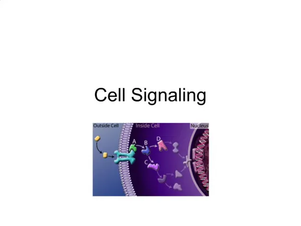
Cell signaling Lecture 8
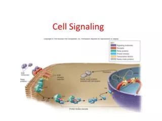
Transforming growth factor (TGFβ) Receptors/Smad pathway TGFβ receptors BMP7 TGFβ1, TGFβ2, TGFβ3 Dpp Inhibins Activins Autocrine/Paracrine signaling Smad proteins located in the cytotsol which moves into the nucleus to regulate transcription
RIII: 280kDa (Monomer, most abundant) RII: 85kDa (Diamer) RI: 55kDa (Diamer)
Ligand binding • Signaling begins with the binding of a ligand to a TGF beta type II receptor. Ligand binding induces formation of complexes containing 2 copies of RI and RII. • The RII is a serine/threonine receptor kinase. • It catalyzes the phosphorylation of the RI. • Each class of ligand binds to a specific RII. • In mammals there are seven known type I receptors and five type II receptors. ATP + [receptor-protein] ADP + [receptor-protein] phosphate
SMAD Phosphorylation • There are five receptor regulated SMADs: SMAD1, SMAD2, SMAD3, SMAD5, and SMAD9. • TGF beta's, Activins, and some GDFs are mediated by SMAD2 and SMAD3, while BMPs,and a few GDFs are mediated by SMAD1, SMAD5 and SMAD9. • The binding of the R-SMAD to the type I receptor is mediated by a zinc double finger FYVE domain containing protein. • Two such proteins that mediate the TGF beta pathway include SARA (The SMAD anchor for receptor activation) and HGS (Hepatocyte growth factor-regulated tyrosine kinase substrate).
SARA recruits an R-SMAD. • SARA permits the binding of the R-SMAD to RI. • SARA orients the R-SMAD such that serine residue on its C-terminus faces the catalytic region of the RI. • The RI phosphorylates the serine residue of the R-SMAD. • Phosphorylation induces a conformational change in the MH2 domain of the R-SMAD and its subsequent dissociation from the receptor complex and SARA.
CoSMAD binding • The phosphorylated R-SMAD has a high affinity for a coSMAD (e.g. SMAD4) and forms a complex with one. • The phosphate group does not act as a docking site for coSMAD, rather the phosphorylation opens up an amino acid stretch allowing interaction.
Transcription • The phosphorylated RSMAD/coSMAD complex enters the nucleus where it binds transcription promoters/cofactors and causes the transcription of DNA. • Bone morphogenetic proteins cause the transcription of mRNAs involved inosteogenesis, neurogenesis, and ventral mesoderm specification. • TGF betas cause the transcription of mRNAs involved in apoptosis, extracellular matrix neogenesis and immunosuppression. It is also involved in G1 arrest in thecell cycle. • Activin causes the transcription of mRNAs involved in gonadal growth, embryo differentiation and placenta formation.
Pathway regulation • The TGF beta signaling pathway is involved in a wide range of cellular process and subsequently is very heavily regulated. • There are a variety of mechanisms where the pathway is modulated either positively or negatively
Role of inhibitory SMADs • There are two other SMADs which complete the SMAD family, the inhibitory SMADs (I-SMADS), SMAD6 and SMAD7 (SnoN and Ski). • They play a key role in the regulation of TGF beta signaling and are involved in negative feedback. • Like other SMADs they have an MH1 and an MH2 domain. • SMAD7 competes with other R-SMADs with RI and prevents their phosphorylation. • It resides in the nucleus and upon TGF beta receptor activation translocates to the cytoplasm where it binds the RI. • SMAD6 binds SMAD4 preventing the binding of R-SMADs with the coSMAD. The levels of I-SMAD increase with TGF beta signaling suggesting that they are downstream targets of TGF-beta signaling.