Download
1 / 16
160 likes | 452 Views
Objectives. Purpose: To compare the Norepinephrine content in visual cortex of REMSD animals v. Control animalsBackground:-Sleep and its possible functions-REM sleep and brain development-LTP and synaptic plasticity-Norepinephrine in sleep and brain maturationMethods:-Sleep Electrode Implantation-REMS deprivation procedure-In Vitro analysis using HPLCResultsConclusion.

E N D
1. Norepinephrine and the Role of REM Sleep in Brain Maturation Erin Johnson
Neuroscience Summer Scholar 2006
Dr. Shaffery�s Lab
2. Objectives Purpose: To compare the Norepinephrine content in visual cortex of REMSD animals v. Control animals
Background:
-Sleep and its possible functions
-REM sleep and brain development
-LTP and synaptic plasticity
-Norepinephrine in sleep and brain maturation
Methods:
-Sleep Electrode Implantation
-REMS deprivation procedure
-In Vitro analysis using HPLC
Results
Conclusion
3. Sleep and its Function Humans spend 1/3 of their life in sleep.
Sleep is critical for almost every physiological and psychological process.
Sleep may play a role in
-restoration and recovery
-energy conservation
-memory consolidation
-brain maturation
Sleep consists of two distinct states: REM sleep and NREM sleep, which alternate in repeating cycles throughout the night.
4. REM Sleep is a Highly Activated State Electrophysiological Characteristics of REMS:
-Wake-like EEG
-Atonia
-Rapid eye movements
Neurochemical Characteristics REMS:
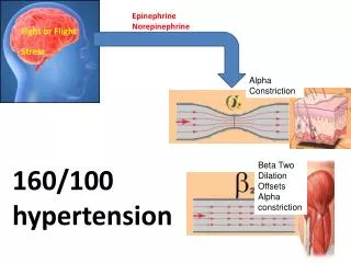
-inhibited aminergic (NE, 5HT) activity
-activated cholinergic (Ach) activity
-inhibited spinal anterior horn cells
5. REM Sleep and Early Development
6. Overview of Plasticity in Rat Visual Cortex Synaptic plasticity is the ability of synapse between two neurons to change in strength.
Long-term Potentiation (LTP) is a model for synaptic plasticity. LTP can be measured as an increase in the amplitude of EPSPs due to a high frequency presynaptic stimulus
Critical period (CP) is the limited time during an organism�s development during which it displays a heightened sensitivity to certain environmental stimuli, and develops in particular ways due to experiences at this time
- CP for rat visual cortex is PN 15 to ~PN 30.
REM sleep provides endogenous cortical stimulation during the CP.
7. Previous Findings Dark-rearing and/or REMS-depriving the animal results in a visual cortex that�s immature compared to normal animals.
REMS-depriving prior the end of CP extends the CP and this developmental-type LTP is maintained.
REMS-depriving after the end of CP up to PN 60 can reopen the CP and unmask mechanisms allowed for developmental LTP.
REMS may be necessary to consolidate the termination of CP.
So what role does norepinephrine play in these phenomena?
8. Norepinephrine, Plasticity, and REMS NE is involved in the developmental plastic changes that occur in the visual cortex during the CP.
REMS may consolidate the CP, while REMSD extends and reopens the CP.
Activity of locus coeruleus (LC) NE is inhibited during REMS, so may be extended during REMSD.
We hypothesize that this extension of LC activation may reduce NE tone in the visual cortex. Evidence has shown that NE is involved in the developmental plastic changes that occur in the visual cortex during the critical period. As I said, further evidence shows that REM sleep may be necessary for the consolidation of these plastic changes during the CP. Seeing that REM sleep depriving the animal somehow extends or reopens the critical period, we wonder what will happen to NE levels in the visual cortex upon REMSD the animals. Since its known that NE cells of the locus coeruleus are inhibited during REM sleep, we proposed that enforced waking by REMSD may cause an over extension of activity of these cells. Accordingly, we hypothesized that this overextension of locus coeruleus NE activity could cause a reduction in the noreadrenergic tone in the visual cortex. In our study used HPLC to measure NE levels in the visual cortex of a REM sleep deprived group compared with a control group, as well as a normal cage-reared group.Evidence has shown that NE is involved in the developmental plastic changes that occur in the visual cortex during the critical period. As I said, further evidence shows that REM sleep may be necessary for the consolidation of these plastic changes during the CP. Seeing that REM sleep depriving the animal somehow extends or reopens the critical period, we wonder what will happen to NE levels in the visual cortex upon REMSD the animals. Since its known that NE cells of the locus coeruleus are inhibited during REM sleep, we proposed that enforced waking by REMSD may cause an over extension of activity of these cells. Accordingly, we hypothesized that this overextension of locus coeruleus NE activity could cause a reduction in the noreadrenergic tone in the visual cortex. In our study used HPLC to measure NE levels in the visual cortex of a REM sleep deprived group compared with a control group, as well as a normal cage-reared group.
9. Rat Experimental Design & Schedule 25-30 34- 35 HOME
CAGE Surgery HOME
CAGE REMS DEP POSTNATAL DAY 0
10. Sleep Electrode Implantation
11. Sleep Recording and Isolation Chamber
12. Sleep Recording and Scoring
13. In Vitro Analysis using HPLC
14. Sleep Scoring Results
15. HPLC Results
16. Conclusion The REMSD was successful and REMS was reduced by 65%.
There was a significant difference in NE in visual cortex between REMSD animals and normals, and almost a significant difference between controls and normals.
NE alone does not seem to be the primary reason for why REM sleep deprivation can extend the CP of developmental synaptic plasticity.
17. Special Thanks TO� Dr. Shaffery
Jorge Lopez
Yillianis Rodrigez
Dr. Regunathan
Dr. Paul
Ronda Richardson
The 2006 Neuroscience Scholars